4IGI

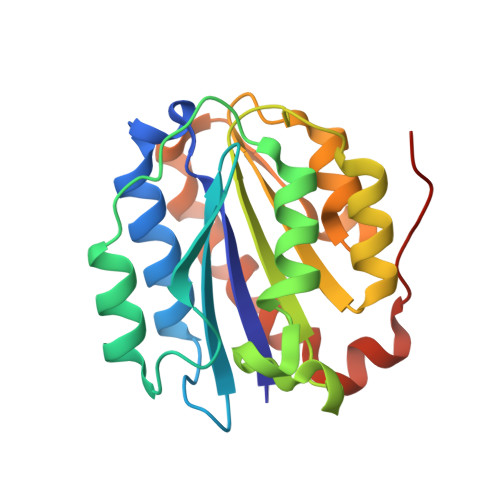
Crystal structure of the Collagen VI alpha3 N5 domain


Experimental Data Snapshot

  • Method: X-RAY DIFFRACTION
  • Resolution: 1.20 Å
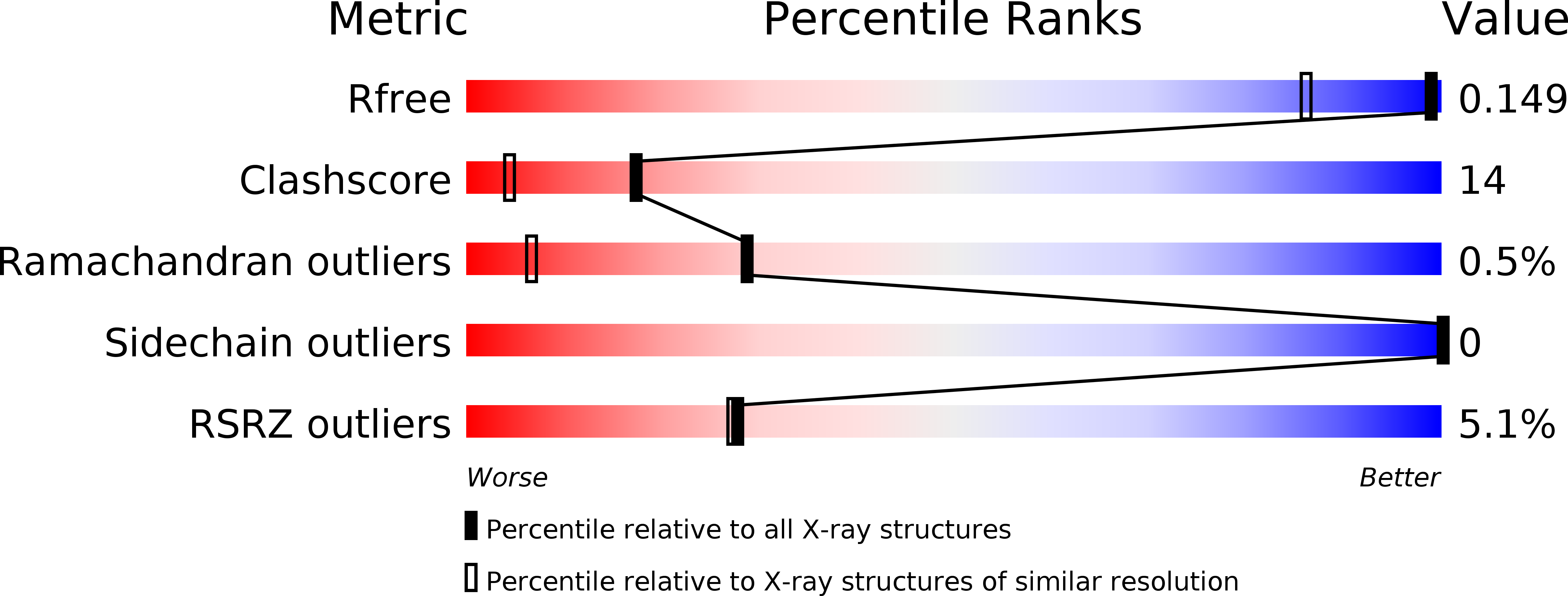
  • R-Value Free: 0.148 
  • R-Value Work: 0.120 
  • R-Value Observed: 0.121 

Starting Model: experimental
View more details

wwPDB Validation   3D Report Full Report


This is version 1.2 of the entry. See complete history


Literature

A structure of a collagen VI VWA domain displays N and C termini at opposite sides of the protein

Becker, A.K.Mikolajek, H.Paulsson, M.Wagener, R.Werner, J.M.

(2014) Structure 22: 199-208

  • DOI: https://doi.org/10.1016/j.str.2013.06.028
  • Primary Citation of Related Structures:  
    4IGI, 4IHK

  • PubMed Abstract: 

    Von Willebrand factor A (VWA) domains are versatile protein interaction domains with N and C termini in close proximity placing spatial constraints on overall protein structure. The 1.2 Å crystal structures of a collagen VI VWA domain and a disease-causing point mutant show C-terminal extensions that place the N and C termini at opposite ends. This allows a "beads-on-a-string" arrangement of multiple VWA domains as observed for ten N-terminal domains of the collagen VI α3 chain. The extension is linked to the core domain by a salt bridge and two hydrophobic patches. Comparison of the wild-type and a muscular dystrophy-associated mutant structure identifies a potential perturbation of a protein interaction interface and indeed, the secretion of mutant collagen VI tetramers is affected. Homology modeling is used to locate a number of disease-associated mutations and analyze their structural impact, which will allow mechanistic analysis of collagen-VI-associated muscular dystrophy phenotypes.


  • Organizational Affiliation

    Center for Biochemistry, Medical Faculty, Center for Molecular Medicine Cologne (CMMC), Cologne Excellence Cluster on Cellular Stress Responses in Aging-Associated Diseases (CECAD), University of Cologne, Cologne 50931, Germany.


Macromolecules
Find similar proteins by:  (by identity cutoff)  |  3D Structure
Entity ID: 1
MoleculeChains Sequence LengthOrganismDetailsImage
Collagen alpha3(VI)203Mus musculusMutation(s): 0 
Gene Names: Col6a3
UniProt & NIH Common Fund Data Resources
Find proteins for E9PWQ3 (Mus musculus)
Explore E9PWQ3 
Go to UniProtKB:  E9PWQ3
IMPC:  MGI:88461
Entity Groups  
Sequence Clusters30% Identity50% Identity70% Identity90% Identity95% Identity100% Identity
UniProt GroupE9PWQ3
Sequence Annotations
Expand
  • Reference Sequence
Experimental Data & Validation

Experimental Data

  • Method: X-RAY DIFFRACTION
  • Resolution: 1.20 Å
  • R-Value Free: 0.148 
  • R-Value Work: 0.120 
  • R-Value Observed: 0.121 
  • Space Group: P 1 21 1
Unit Cell:
Length ( Å )Angle ( ˚ )
a = 37.73α = 90
b = 58.46β = 112.97
c = 39.16γ = 90
Software Package:
Software NamePurpose
HKL-2000data collection
PHASERphasing
PHENIXrefinement
MOSFLMdata reduction
SCALAdata scaling

Structure Validation

View Full Validation Report


Entry History 

Deposition Data

Revision History  (Full details and data files)

  • Version 1.0: 2013-12-18
    Type: Initial release
  • Version 1.1: 2018-12-05
    Changes: Data collection, Database references
  • Version 1.2: 2023-11-08
    Changes: Data collection, Database references, Derived calculations, Refinement description