4IA4 | pdb_00004ia4

Structure of the spinach aquaporin SoPIP2;1 at pH 6


Experimental Data Snapshot

  • Method: X-RAY DIFFRACTION
  • Resolution: 3.10 Å
  • R-Value Free: 
    0.243 (Depositor), 0.244 (DCC) 
  • R-Value Work: 
    0.237 (Depositor), 0.233 (DCC) 
  • R-Value Observed: 
    0.237 (Depositor) 

Starting Model: experimental
View more details

wwPDB Validation 3D Report Full Report

Validation slider image for 4IA4

This is version 1.2 of the entry. See complete history

Literature

Structural basis for pH gating of plant aquaporins

Frick, A.Jarva, M.Tornroth-Horsefield, S.

(2013) FEBS Lett 587: 989-993

  • DOI: https://doi.org/10.1016/j.febslet.2013.02.038
  • Primary Citation Related Structures: 
    4IA4

  • PubMed Abstract: 

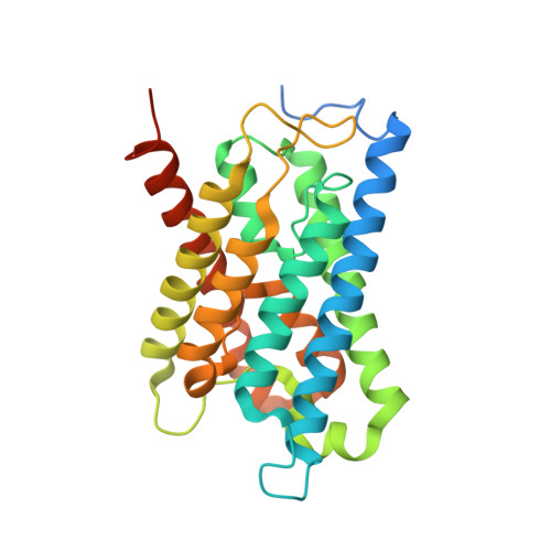
    Plants have evolved to cope with fluctuations in water supply by gating their water channels known as aquaporins. During flooding, a rapid drop of cytosolic pH due to anoxia leads to a simultaneous closure of the aquaporins in the plasma membrane. The closing mechanism has been suggested to involve a conserved histidine on cytosolic loop D. Here we report the crystal structure of a spinach aquaporin at low pH, revealing for the first time the structural basis for how this pH-sensitive histidine helps to keep the aquaporin in a closed state.


  • Organizational Affiliation
    • Department of Chemistry and Molecular Biology, University of Gothenburg, Gothenburg, Sweden.

Macromolecule Content 

  • Total Structure Weight: 122.13 kDa 
  • Atom Count: 7,290 
  • Modeled Residue Count: 967 
  • Deposited Residue Count: 1,124 
  • Unique protein chains: 1

Macromolecules

Find similar proteins by:|  3D Structure
Entity ID: 1
MoleculeChains  Sequence LengthOrganismDetailsImage
Aquaporin
A, B, C, D
281Spinacia oleraceaMutation(s): 0 
Membrane Entity: Yes 
UniProt
Find proteins for Q41372 (Spinacia oleracea)
Explore Q41372 
Go to UniProtKB:  Q41372
Entity Groups
Sequence Clusters30% Identity50% Identity70% Identity90% Identity95% Identity100% Identity
UniProt GroupQ41372
Sequence Annotations
Expand
Reference Sequence

Small Molecules

Ligands 1 Unique
IDChains Name / Formula / InChI Key2D Diagram3D Interactions
HG

Query on HG


Download:Ideal Coordinates CCD File
E [auth A]
F [auth A]
G [auth A]
H [auth A]
I [auth B]
E [auth A],
F [auth A],
G [auth A],
H [auth A],
I [auth B],
J [auth B],
K [auth B],
L [auth B],
M [auth C],
N [auth C],
O [auth D],
P [auth D]
MERCURY (II) ION
Hg
BQPIGGFYSBELGY-UHFFFAOYSA-N

Experimental Data & Validation

Experimental Data

  • Method: X-RAY DIFFRACTION
  • Resolution: 3.10 Å
  • R-Value Free:  0.243 (Depositor), 0.244 (DCC) 
  • R-Value Work:  0.237 (Depositor), 0.233 (DCC) 
  • R-Value Observed: 0.237 (Depositor) 
Space Group: P 21 21 2
Unit Cell:
Length ( Å )Angle ( ˚ )
a = 178.227α = 90
b = 104.19β = 90
c = 66.523γ = 90
Software Package:
Software NamePurpose
ADSCdata collection
MOLREPphasing
CNSrefinement
MOSFLMdata reduction
SCALAdata scaling

Structure Validation

View Full Validation Report


Entry History 

Deposition Data

Revision History  (Full details and data files)

  • Version 1.0: 2013-05-29
    Type: Initial release
  • Version 1.1: 2023-11-08
    Changes: Data collection, Database references, Derived calculations, Refinement description
  • Version 1.2: 2024-11-13
    Changes: Structure summary