4I92 | pdb_00004i92

Structure of the BSK8 kinase domain


Experimental Data Snapshot

  • Method: X-RAY DIFFRACTION
  • Resolution: 1.60 Å
  • R-Value Free: 
    0.183 (Depositor), 0.203 (DCC) 
  • R-Value Work: 
    0.160 (Depositor) 
  • R-Value Observed: 
    0.161 (Depositor) 

wwPDB Validation 3D Report Full Report

Validation slider image for 4I92

This is version 1.2 of the entry. See complete history

Literature

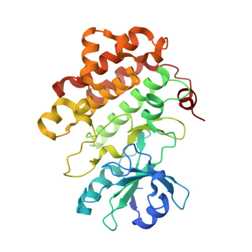
Structural Characterization of the RLCK Family Member BSK8: A Pseudokinase with an Unprecedented Architecture

Grutter, C.Sreeramulu, S.Sessa, G.Rauh, D.

(2013) J Mol Biology 425: 4455-4467

  • DOI: https://doi.org/10.1016/j.jmb.2013.07.034
  • Primary Citation Related Structures: 
    4I92, 4I93, 4I94

  • PubMed Abstract: 

    Brassinosteroid signaling kinases (BSKs) are plant-specific receptor-like cytoplasmic protein kinases involved in the brassinosteroid signaling pathway. Unlike common protein kinases, they possess a naturally occurring alanine residue at the "gatekeeper" position, as well as other sequence variations. How BSKs activate downstream proteins such as BSU1, as well as the structural consequences of their unusual sequential features, was unclear. We crystallized the catalytic domain of BSK8 and solved its structure by multiple-wavelength anomalous dispersion phasing methods to a resolution of 1.5Å. In addition, a co-crystal structure of BSK8 with 5-adenylyl imidodiphosphate (AMP-PNP) revealed unusual conformational arrangements of the nucleotide phosphate groups and catalytic key motifs, typically not observed for active protein kinases. Sequential analysis and comparisons with known pseudokinase structures suggest that BSKs represent constitutively inactive protein kinases that regulate brassinosteroid signal transfer through an allosteric mechanism.


  • Organizational Affiliation
    • Technische Universität Dortmund Fakultät Chemie und Chemische Biologie, Otto-Hahn-Strasse 6, D-44227 Dortmund, Germany.

Macromolecule Content 

  • Total Structure Weight: 34.42 kDa 
  • Atom Count: 2,622 
  • Modeled Residue Count: 289 
  • Deposited Residue Count: 300 
  • Unique protein chains: 1

Macromolecules

Find similar proteins by:|  3D Structure
Entity ID: 1
MoleculeChains  Sequence LengthOrganismDetailsImage
Probable serine/threonine-protein kinase At5g41260300Arabidopsis thalianaMutation(s): 0 
Gene Names: At5g41260K1O13.5
EC: 2.7.11.1
UniProt
Find proteins for Q9FHD7 (Arabidopsis thaliana)
Explore Q9FHD7 
Go to UniProtKB:  Q9FHD7
Entity Groups
Sequence Clusters30% Identity50% Identity70% Identity90% Identity95% Identity100% Identity
UniProt GroupQ9FHD7
Sequence Annotations
Expand
Reference Sequence

Small Molecules

Ligands 1 Unique
IDChains Name / Formula / InChI Key2D Diagram3D Interactions
CL

Query on CL


Download:Ideal Coordinates CCD File
B [auth A]CHLORIDE ION
Cl
VEXZGXHMUGYJMC-UHFFFAOYSA-M

Experimental Data & Validation

Experimental Data

  • Method: X-RAY DIFFRACTION
  • Resolution: 1.60 Å
  • R-Value Free:  0.183 (Depositor), 0.203 (DCC) 
  • R-Value Work:  0.160 (Depositor) 
  • R-Value Observed: 0.161 (Depositor) 
Space Group: P 1 21 1
Unit Cell:
Length ( Å )Angle ( ˚ )
a = 35.09α = 90
b = 76.99β = 104.86
c = 50.57γ = 90
Software Package:
Software NamePurpose
XSCALEdata scaling
PHASERphasing
REFMACrefinement
PDB_EXTRACTdata extraction
XDSdata scaling
XDSdata reduction

Structure Validation

View Full Validation Report


Entry History 

Deposition Data

Revision History  (Full details and data files)

  • Version 1.0: 2013-08-14
    Type: Initial release
  • Version 1.1: 2013-11-20
    Changes: Database references
  • Version 1.2: 2024-03-20
    Changes: Data collection, Database references, Derived calculations