4I5E | pdb_00004i5e

Crystal structure of Ralstonia sp. alcohol dehydrogenase in complex with NADP+


Experimental Data Snapshot

  • Method: X-RAY DIFFRACTION
  • Resolution: 2.80 Å
  • R-Value Free: 
    0.305 (Depositor), 0.312 (DCC) 
  • R-Value Work: 
    0.261 (Depositor), 0.268 (DCC) 
  • R-Value Observed: 
    0.263 (Depositor) 

Starting Model: experimental
View more details

wwPDB Validation 3D Report Full Report

Validation slider image for 4I5E

Ligand Structure Quality Assessment 


This is version 1.2 of the entry. See complete history

Literature

Crystallographic analysis and structure-guided engineering of NADPH-dependent Ralstonia sp. Alcohol dehydrogenase toward NADH cosubstrate specificity.

Lerchner, A.Jarasch, A.Meining, W.Schiefner, A.Skerra, A.

(2013) Biotechnol Bioeng 110: 2803-2814

  • DOI: https://doi.org/10.1002/bit.24956
  • Primary Citation Related Structures: 
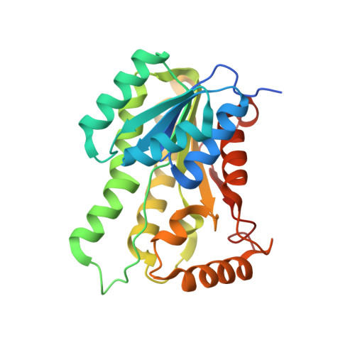
    4I5D, 4I5E, 4I5F, 4I5G

  • PubMed Abstract: 

    The NADP⁺-dependent alcohol dehydrogenase from Ralstonia sp. (RasADH) belongs to the protein superfamily of short-chain dehydrogenases/reductases (SDRs). As an enzyme that accepts different types of substrates--including bulky-bulky as well as small-bulky secondary alcohols or ketones--with high stereoselectivity, it offers potential as a biocatalyst for industrial biotechnology. To understand substrate and cosubstrate specificities of RasADH we determined the crystal structure of the apo-enzyme as well as its NADP⁺-bound state with resolutions down to 2.8 Å. RasADH displays a homotetrameric quaternary structure that can be described as a dimer of homodimers while in each subunit a seven-stranded parallel β-sheet, flanked by three α-helices on each side, forms a Rossmann fold-type dinucleotide binding domain. Docking of the well-known substrate (S)-1-phenylethanol clearly revealed the structural determinants of stereospecificity. To favor practical RasADH application in the context of established cofactor recycling systems, for example, those involving an NADH-dependent amino acid dehydrogenase, we attempted to rationally change its cosubstrate specificity from NADP⁺ to NAD⁺ utilizing the structural information that NADP⁺ specificity is largely governed by the residues Asn15, Gly37, Arg38, and Arg39. Furthermore, an extensive sequence alignment with homologous dehydrogenases that have different cosubstrate specificities revealed a modified general SDR motif ASNG (instead of NNAG) at positions 86-89 of RasADH. Consequently, we constructed mutant enzymes with one (G37D), four (N15G/G37D/R38V/R39S), and six (N15G/G37D/R38V/R39S/A86N/S88A) amino acid exchanges. RasADH (N15G/G37D/R38V/R39S) was better able to accept NAD⁺ while showing much reduced catalytic efficiency with NADP⁺, leading to a change in NADH/NADPH specificity by a factor of ∼3.6 million.


  • Organizational Affiliation
    • Munich Center for Integrated Protein Science, CIPS-M, and Lehrstuhl für Biologische Chemie, Technische Universität München, 85350, Freising-Weihenstephan, Germany.

Macromolecule Content 

  • Total Structure Weight: 231.74 kDa 
  • Atom Count: 15,560 
  • Modeled Residue Count: 1,992 
  • Deposited Residue Count: 2,096 
  • Unique protein chains: 1

Macromolecules

Find similar proteins by:|  3D Structure
Entity ID: 1
MoleculeChains  Sequence LengthOrganismDetailsImage
Alclohol dehydrogenase/short-chain dehydrogenase
A, B, C, D, E
A, B, C, D, E, F, G, H
262Ralstonia sp. DSMZ 6428Mutation(s): 0 
Gene Names: RasADH
UniProt
Find proteins for C0IR58 (Ralstonia sp. DSMZ 6428)
Explore C0IR58 
Go to UniProtKB:  C0IR58
Entity Groups
Sequence Clusters30% Identity50% Identity70% Identity90% Identity95% Identity100% Identity
UniProt GroupC0IR58
Sequence Annotations
Expand
Reference Sequence

Small Molecules

Ligands 2 Unique
IDChains Name / Formula / InChI Key2D Diagram3D Interactions
NAP

Query on NAP


Download:Ideal Coordinates CCD File
I [auth A]
K [auth B]
M [auth C]
O [auth D]
P [auth E]
I [auth A],
K [auth B],
M [auth C],
O [auth D],
P [auth E],
R [auth F],
S [auth G],
T [auth H]
NADP NICOTINAMIDE-ADENINE-DINUCLEOTIDE PHOSPHATE
C21 H28 N7 O17 P3
XJLXINKUBYWONI-NNYOXOHSSA-N
GOL

Query on GOL


Download:Ideal Coordinates CCD File
J [auth A],
L [auth B],
N [auth C],
Q [auth E]
GLYCEROL
C3 H8 O3
PEDCQBHIVMGVHV-UHFFFAOYSA-N

Experimental Data & Validation

Experimental Data

  • Method: X-RAY DIFFRACTION
  • Resolution: 2.80 Å
  • R-Value Free:  0.305 (Depositor), 0.312 (DCC) 
  • R-Value Work:  0.261 (Depositor), 0.268 (DCC) 
  • R-Value Observed: 0.263 (Depositor) 
Space Group: P 1
Unit Cell:
Length ( Å )Angle ( ˚ )
a = 73.342α = 80.98
b = 74.549β = 85.99
c = 132.689γ = 64.6
Software Package:
Software NamePurpose
XSCALEdata scaling
REFMACrefinement
PDB_EXTRACTdata extraction
MAR345dtbdata collection
XDSdata reduction
PHASERphasing

Structure Validation

View Full Validation Report


Ligand Structure Quality Assessment 


Entry History 

Deposition Data

Revision History  (Full details and data files)

  • Version 1.0: 2013-06-12
    Type: Initial release
  • Version 1.1: 2013-10-09
    Changes: Database references
  • Version 1.2: 2023-09-20
    Changes: Data collection, Database references, Derived calculations, Refinement description