4I25 | pdb_00004i25

2.00 Angstroms X-ray crystal structure of NAD- and substrate-bound 2-aminomuconate 6-semialdehyde dehydrogenase from Pseudomonas fluorescens


Experimental Data Snapshot

  • Method: X-RAY DIFFRACTION
  • Resolution: 2.00 Å
  • R-Value Free: 
    0.208 (Depositor), 0.211 (DCC) 
  • R-Value Work: 
    0.162 (Depositor), 0.168 (DCC) 
  • R-Value Observed: 
    0.165 (Depositor) 

Starting Model: experimental
View more details

wwPDB Validation 3D Report Full Report

Validation slider image for 4I25

Ligand Structure Quality Assessment 


This is version 1.3 of the entry. See complete history

Literature

Crystallographic and spectroscopic snapshots reveal a dehydrogenase in action.

Huo, L.Davis, I.Liu, F.Andi, B.Esaki, S.Iwaki, H.Hasegawa, Y.Orville, A.M.Liu, A.

(2015) Nat Commun 6: 5935-5935

  • DOI: https://doi.org/10.1038/ncomms6935
  • Primary Citation Related Structures: 
    4I1W, 4I25, 4I26, 4I2R, 4NPI, 4OE2, 4OU2, 4OUB

  • PubMed Abstract: 

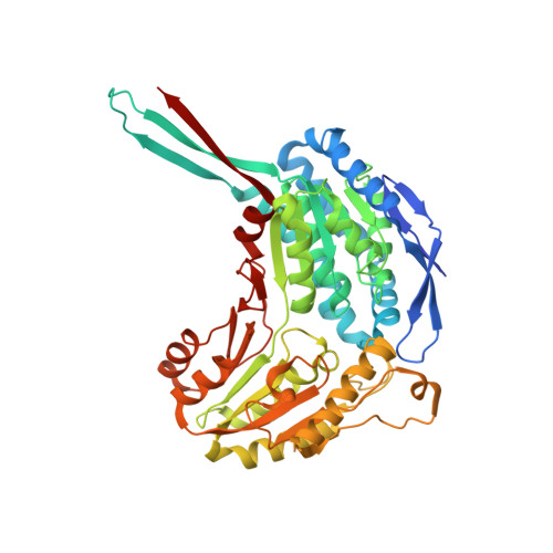
    Aldehydes are ubiquitous intermediates in metabolic pathways and their innate reactivity can often make them quite unstable. There are several aldehydic intermediates in the metabolic pathway for tryptophan degradation that can decay into neuroactive compounds that have been associated with numerous neurological diseases. An enzyme of this pathway, 2-aminomuconate-6-semialdehyde dehydrogenase, is responsible for 'disarming' the final aldehydic intermediate. Here we show the crystal structures of a bacterial analogue enzyme in five catalytically relevant forms: resting state, one binary and two ternary complexes, and a covalent, thioacyl intermediate. We also report the crystal structures of a tetrahedral, thiohemiacetal intermediate, a thioacyl intermediate and an NAD(+)-bound complex from an active site mutant. These covalent intermediates are characterized by single-crystal and solution-state electronic absorption spectroscopy. The crystal structures reveal that the substrate undergoes an E/Z isomerization at the enzyme active site before an sp(3)-to-sp(2) transition during enzyme-mediated oxidation.


  • Organizational Affiliation
    • 1] Department of Chemistry, Georgia State University, Atlanta, Georgia 30303, USA [2] Molecular Basis of Disease Area of Focus Program, Georgia State University, Atlanta, Georgia 30303, USA.

Macromolecule Content 

  • Total Structure Weight: 219.21 kDa 
  • Atom Count: 16,301 
  • Modeled Residue Count: 1,932 
  • Deposited Residue Count: 2,000 
  • Unique protein chains: 1

Macromolecules

Find similar proteins by:|  3D Structure
Entity ID: 1
MoleculeChains  Sequence LengthOrganismDetailsImage
2-aminomuconate 6-semialdehyde dehydrogenase
A, B, C, D
500Pseudomonas fluorescensMutation(s): 0 
Gene Names: nbaE
UniProt
Find proteins for Q83V33 (Pseudomonas fluorescens)
Explore Q83V33 
Go to UniProtKB:  Q83V33
Entity Groups
Sequence Clusters30% Identity50% Identity70% Identity90% Identity95% Identity100% Identity
UniProt GroupQ83V33
Sequence Annotations
Expand
Reference Sequence

Small Molecules

Ligands 3 Unique
IDChains Name / Formula / InChI Key2D Diagram3D Interactions
NAD

Query on NAD


Download:Ideal Coordinates CCD File
E [auth A],
J [auth B],
K [auth C],
N [auth D]
NICOTINAMIDE-ADENINE-DINUCLEOTIDE
C21 H27 N7 O14 P2
BAWFJGJZGIEFAR-NNYOXOHSSA-N
6OD

Query on 6OD


Download:Ideal Coordinates CCD File
G [auth A],
I [auth B],
M [auth C],
P [auth D]
(2E,4E)-2-amino-6-oxohexa-2,4-dienoic acid
C6 H7 N O3
QCGTZPZKJPTAEP-NRNIAZNESA-N
NA

Query on NA


Download:Ideal Coordinates CCD File
F [auth A],
H [auth B],
L [auth C],
O [auth D]
SODIUM ION
Na
FKNQFGJONOIPTF-UHFFFAOYSA-N

Experimental Data & Validation

Experimental Data

  • Method: X-RAY DIFFRACTION
  • Resolution: 2.00 Å
  • R-Value Free:  0.208 (Depositor), 0.211 (DCC) 
  • R-Value Work:  0.162 (Depositor), 0.168 (DCC) 
  • R-Value Observed: 0.165 (Depositor) 
Space Group: P 21 21 21
Unit Cell:
Length ( Å )Angle ( ˚ )
a = 88.253α = 90
b = 142.932β = 90
c = 175.125γ = 90
Software Package:
Software NamePurpose
PHENIXrefinement
PDB_EXTRACTdata extraction
SERGUIdata collection
HKL-2000data reduction
SCALEPACKdata scaling
PHENIXphasing

Structure Validation

View Full Validation Report


Ligand Structure Quality Assessment 


Entry History 

Deposition Data

Revision History  (Full details and data files)

  • Version 1.0: 2014-05-21
    Type: Initial release
  • Version 1.1: 2015-01-21
    Changes: Database references
  • Version 1.2: 2017-11-15
    Changes: Refinement description
  • Version 1.3: 2023-09-20
    Changes: Data collection, Database references, Derived calculations, Refinement description