4HXV | pdb_00004hxv

Crystal structure of 3'(2'),5'-bisphosphate nucleotidase1 from Entamoeba histolytica in complex with AMP and metal ions


Experimental Data Snapshot

  • Method: X-RAY DIFFRACTION
  • Resolution: 2.60 Å
  • R-Value Free: 
    0.283 (Depositor), 0.280 (DCC) 
  • R-Value Work: 
    0.230 (Depositor), 0.229 (DCC) 
  • R-Value Observed: 
    0.233 (Depositor) 

Starting Model: experimental
View more details

wwPDB Validation 3D Report Full Report

Validation slider image for 4HXV

Ligand Structure Quality Assessment 


This is version 1.2 of the entry. See complete history

Literature

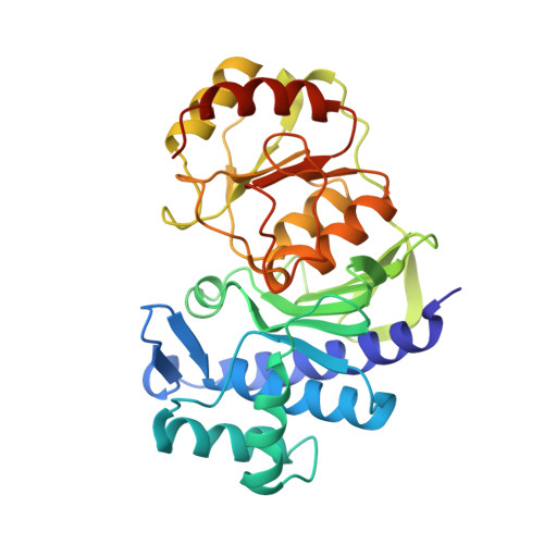
Structural elucidation of a dual-activity PAP phosphatase-1 from Entamoeba histolytica capable of hydrolysing both 3'-phosphoadenosine 5'-phosphate and inositol 1,4-bisphosphate

Faisal Tarique, K.Arif Abdul Rehman, S.Gourinath, S.

(2014) Acta Crystallogr D Biol Crystallogr 70: 2019-2031

  • DOI: https://doi.org/10.1107/S1399004714010268
  • Primary Citation Related Structures: 
    4HXV, 4O7I

  • PubMed Abstract: 

    The enzyme 3'-phosphoadenosine 5'-phosphatase-1 (PAP phosphatase-1) is a member of the Li(+)-sensitive Mg(2+)-dependent phosphatase superfamily, or inositol monophosphatase (IMPase) superfamily, and is an important regulator of the sulfate-activation pathway in all living organisms. Inhibition of this enzyme leads to accumulation of the toxic byproduct 3'-phosphoadenosine 5'-phosphate (PAP), which could be lethal to the organism. Genomic analysis of Entamoeba histolytica suggests the presence of two isoforms of PAP phosphatase. The PAP phosphatase-1 isoform of this organism is shown to be active over wide ranges of pH and temperature. Interestingly, this enzyme is inhibited by submillimolar concentrations of Li(+), while being insensitive to Na(+). Interestingly, the enzyme showed activity towards both PAP and inositol 1,4-bisphosphate and behaved as an inositol polyphosphate 1-phosphatase. Crystal structures of this enzyme in its native form and in complex with adenosine 5'-monophosphate have been determined to 2.1 and 2.6 Å resolution, respectively. The PAP phosphatase-1 structure is divided into two domains, namely α+β and α/β, and the substrate and metal ions bind between them. This is a first structure of any PAP phosphatase to be determined from a human parasitic protozoan. This enzyme appears to function using a mechanism involving three-metal-ion assisted catalysis. Comparison with other structures indicates that the sensitivity to alkali-metal ions may depend on the orientation of a specific catalytic loop.


  • Organizational Affiliation
    • School of Life Sciences, Jawaharlal Nehru University, New Delhi 110 067, India.

Macromolecule Content 

  • Total Structure Weight: 36.55 kDa 
  • Atom Count: 2,532 
  • Modeled Residue Count: 317 
  • Deposited Residue Count: 325 
  • Unique protein chains: 1

Macromolecules

Find similar proteins by:|  3D Structure
Entity ID: 1
MoleculeChains  Sequence LengthOrganismDetailsImage
3'(2'),5'-bisphosphate nucleotidase, putative325Entamoeba histolyticaMutation(s): 0 
Gene Names: EHI_193350
EC: 3.1.3.7 (PDB Primary Data), 3.1.3.57 (UniProt)
UniProt
Find proteins for C4M4T9 (Entamoeba histolytica (strain ATCC 30459 / HM-1:IMSS / ABRM))
Explore C4M4T9 
Go to UniProtKB:  C4M4T9
Entity Groups
Sequence Clusters30% Identity50% Identity70% Identity90% Identity95% Identity100% Identity
UniProt GroupC4M4T9
Sequence Annotations
Expand
Reference Sequence

Experimental Data & Validation

Experimental Data

  • Method: X-RAY DIFFRACTION
  • Resolution: 2.60 Å
  • R-Value Free:  0.283 (Depositor), 0.280 (DCC) 
  • R-Value Work:  0.230 (Depositor), 0.229 (DCC) 
  • R-Value Observed: 0.233 (Depositor) 
Space Group: P 21 21 21
Unit Cell:
Length ( Å )Angle ( ˚ )
a = 39.618α = 90
b = 63.877β = 90
c = 117.845γ = 90
Software Package:
Software NamePurpose
MAR345dtbdata collection
PHASERphasing
REFMACrefinement
HKL-2000data reduction
HKL-2000data scaling

Structure Validation

View Full Validation Report


Ligand Structure Quality Assessment 


Entry History 

Deposition Data

Revision History  (Full details and data files)

  • Version 1.0: 2013-11-13
    Type: Initial release
  • Version 1.1: 2014-08-06
    Changes: Database references
  • Version 1.2: 2023-11-08
    Changes: Data collection, Database references, Derived calculations, Refinement description