4HVU | pdb_00004hvu

Crystal structure of the T98D c-Src-SH3 domain mutant in complex with the high affinity peptide APP12


Experimental Data Snapshot

  • Method: X-RAY DIFFRACTION
  • Resolution: 0.98 Å
  • R-Value Free: 
    0.160 (Depositor) 
  • R-Value Work: 
    0.145 (Depositor) 
  • R-Value Observed: 
    0.146 (Depositor) 

Starting Model: experimental
View more details

wwPDB Validation 3D Report Full Report

Validation slider image for 4HVU

This is version 1.2 of the entry. See complete history

Literature

Atomic resolution structures of the c-Src SH3 domain in complex with two high-affinity peptides from classes I and II.

Bacarizo, J.Camara-Artigas, A.

(2013) Acta Crystallogr D Biol Crystallogr 69: 756-766

  • DOI: https://doi.org/10.1107/S0907444913001522
  • Primary Citation Related Structures: 
    4HVU, 4HVV, 4HVW

  • PubMed Abstract: 

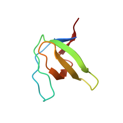
    The atomic resolution crystal structures of complexes between the SH3 domain of the c-Src tyrosine kinase and two high-affinity peptides belonging to class I and class II have been solved. The crystals of the Thr98Asp and Thr98Glu mutants in complex with the APP12 peptide (APPLPPRNRPRL) belonged to the trigonal space group P3121 and in both cases the asymmetric unit was composed of one molecule of the SH3-APP12 complex. The crystals of the Thr98Glu mutant in complex with the VSL12 peptide (VSLARRPLPLP) belonged to the trigonal space group P3221 and the asymmetric unit was also composed of a single molecule of the SH3-VSL12 complex. All crystals were obtained in the presence of PEG 300 under the same conditions as reported for the intertwined dimeric structure of the c-Src SH3 domain, but the presence of the peptide stabilizes the monomeric form of the domain. These structures allow a detailed analysis of the role of salt bridges, cation-π interactions and hydrogen bonds in the binding of proline-rich motifs to the c-Src SH3 domain. Moreover, these crystallographic structures allow the role of water molecules in the binding of these motifs to the c-Src SH3 domain to be studied for the first time.


  • Organizational Affiliation
    • Department of Chemistry and Physics, University of Almería, Agrifood Campus of International Excellence, Carretera de Sacramento, 04120 Almería, Spain.

Macromolecule Content 

  • Total Structure Weight: 8.28 kDa 
  • Atom Count: 650 
  • Modeled Residue Count: 68 
  • Deposited Residue Count: 72 
  • Unique protein chains: 2

Macromolecules

Find similar proteins by:|  3D Structure
Entity ID: 1
MoleculeChains  Sequence LengthOrganismDetailsImage
Proto-oncogene tyrosine-protein kinase Src61Gallus gallusMutation(s): 2 
Gene Names: SRC
EC: 2.7.10.2
UniProt
Find proteins for P00523 (Gallus gallus)
Explore P00523 
Go to UniProtKB:  P00523
Entity Groups
Sequence Clusters30% Identity50% Identity70% Identity90% Identity95% Identity100% Identity
UniProt GroupP00523
Sequence Annotations
Expand
Reference Sequence
Find similar proteins by:  Sequence   |   3D Structure  
Entity ID: 2
MoleculeChains  Sequence LengthOrganismDetailsImage
SYNTHETIC PEPTIDE Acetyl-APPLPPRNRP11N/AMutation(s): 0 

Experimental Data & Validation

Experimental Data

  • Method: X-RAY DIFFRACTION
  • Resolution: 0.98 Å
  • R-Value Free:  0.160 (Depositor) 
  • R-Value Work:  0.145 (Depositor) 
  • R-Value Observed: 0.146 (Depositor) 
Space Group: P 31 2 1
Unit Cell:
Length ( Å )Angle ( ˚ )
a = 31.503α = 90
b = 31.503β = 90
c = 106.71γ = 120
Software Package:
Software NamePurpose
Aimlessdata scaling
PHENIXrefinement
PDB_EXTRACTdata extraction
ADSCdata collection
XDSdata reduction
PHASERphasing

Structure Validation

View Full Validation Report


Entry History 

Deposition Data

Revision History  (Full details and data files)

  • Version 1.0: 2013-05-01
    Type: Initial release
  • Version 1.1: 2013-06-19
    Changes: Database references
  • Version 1.2: 2023-09-20
    Changes: Data collection, Database references, Derived calculations, Refinement description