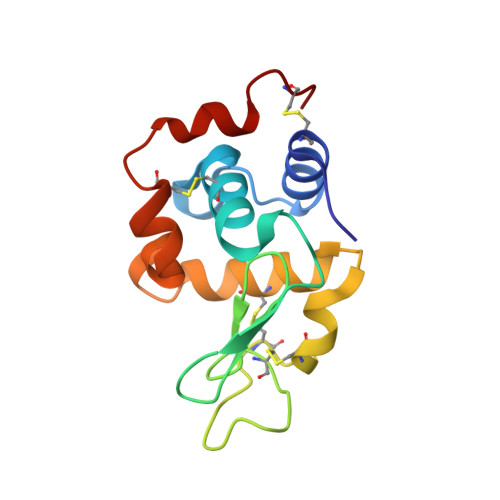
Mitigation of X-ray damage in macromolecular crystallography by submicrometre line focusing.
Finfrock, Y.Z., Stern, E.A., Alkire, R.W., Kas, J.J., Evans-Lutterodt, K., Stein, A., Duke, N., Lazarski, K., Joachimiak, A.(2013) Acta Crystallogr D Biol Crystallogr 69: 1463-1469
- PubMed: 23897469
- DOI: https://doi.org/10.1107/S0907444913009335
- Primary Citation of Related Structures:
4HTK, 4HTN, 4HTQ - PubMed Abstract:
Reported here are measurements of the penetration depth and spatial distribution of photoelectron (PE) damage excited by 18.6 keV X-ray photons in a lysozyme crystal with a vertical submicrometre line-focus beam of 0.7 µm full-width half-maximum (FWHM). The experimental results determined that the penetration depth of PEs is 5 ± 0.5 µm with a monotonically decreasing spatial distribution shape, resulting in mitigation of diffraction signal damage. This does not agree with previous theoretical predication that the mitigation of damage requires a peak of damage outside the focus. A new improved calculation provides some qualitative agreement with the experimental results, but significant errors still remain. The mitigation of radiation damage by line focusing was measured experimentally by comparing the damage in the X-ray-irradiated regions of the submicrometre focus with the large-beam case under conditions of equal exposure and equal volumes of the protein crystal, and a mitigation factor of 4.4 ± 0.4 was determined. The mitigation of radiation damage is caused by spatial separation of the dominant PE radiation-damage component from the crystal region of the line-focus beam that contributes the diffraction signal. The diffraction signal is generated by coherent scattering of incident X-rays (which introduces no damage), while the overwhelming proportion of damage is caused by PE emission as X-ray photons are absorbed.
Organizational Affiliation:
Physics Department, University of Washington, Seattle, WA 98195, USA.


