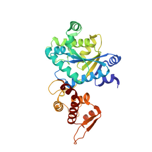
Crystal structure of Tyrosine-tRNA ligase mutant complexed with unnatural amino acid 3-o-methyl-Tyrosine
Yu, Y., Zhou, Q., Dong, J., Li, J., Xiaoxuan, L., Mukherjee, A., Ouyang, H., Nilges, M., Li, H., Gao, F., Gong, W., Lu, Y., Wang, J.To be published.