4HPB | pdb_00004hpb

Crystal structure of Nitrophorin 4 from Rhodnius prolixus Complexed with Beta-Mercaptoethanol at pH 7.4


Experimental Data Snapshot

  • Method: X-RAY DIFFRACTION
  • Resolution: 1.60 Å
  • R-Value Free: 
    0.179 (Depositor), 0.177 (DCC) 
  • R-Value Work: 
    0.142 (Depositor), 0.136 (DCC) 
  • R-Value Observed: 
    0.144 (Depositor) 

Starting Model: experimental
View more details

wwPDB Validation 3D Report Full Report

Validation slider image for 4HPB

Ligand Structure Quality Assessment 


This is version 1.2 of the entry. See complete history

Literature

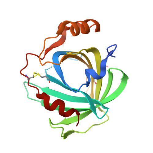
Complexes of ferriheme nitrophorin 4 with low-molecular weight thiol(ate)s occurring in blood plasma

He, C.Nishikawa, K.Erdem, O.F.Reijerse, E.Ogata, H.Lubitz, W.Knipp, M.

(2013) J Inorg Biochem 122: 38-48

  • DOI: https://doi.org/10.1016/j.jinorgbio.2013.01.012
  • Primary Citation Related Structures: 
    4HPA, 4HPB, 4HPC, 4HPD

  • PubMed Abstract: 

    Nitrophorins are proteins occurring in the saliva of the blood-sucking insect Rhodnius prolixus to carry NO as a vasodilator and blood-coagulation inhibitor into the victim's tissue. It was suggested that the rate of NO release can be enhanced by the blood-plasma component L-cysteine [J.M.C.Ribeiro, Insect Biochem. Mol. Biol. 26 (1996) 899-905]. However, the mechanism of the reaction is not clear. In the attempt to exploit the reaction in detail, complexes of nitrophorin 4 (NP4) with the thiols 2-mercaptoethanol, L-cysteine, and L-homocysteine and with HS(-) were formed and characterized under anaerobic conditions using absorption spectroscopy, X-ray crystallography, and EPR spectroscopy. In contrast to met-myoglobin, which is reduced by L-cysteine, all four compounds form low-spin Fe(III) complexes with NP4. The weak equilibration constants (167-5200 M(-1)) neither support significant complexation nor the simple displacement of NO in vivo. Both amino acid based thiols form additional H-bonds with side chains of the heme pocket entry. Glutathione and L-methionine did not form a complex, indicating the specificity of the complexes with L-cysteine and L-homocysteine. Continuous wave EPR spectroscopy reveals the simultaneous existence of three low-spin systems in each case that are attributed to various protonation and/or conformational stages in the heme pocket. Electron nuclear double resonance (ENDOR) spectroscopy demonstrates that the thiol sulfurs are, at least in part, protonated. Overall, the results not only demonstrate the good accessibility of the NP4 heme center by biologically relevant thiols, but also represent the first structural characterization of a ferriheme protein in complex with L-cysteine L-homocysteine.


  • Organizational Affiliation
    • Max-Planck-Institut für Chemische Energiekonversion, Stiftstrasse 34-36, D-45470 Mülheim an der Ruhr, Germany.

Macromolecule Content 

  • Total Structure Weight: 20.99 kDa 
  • Atom Count: 1,651 
  • Modeled Residue Count: 179 
  • Deposited Residue Count: 184 
  • Unique protein chains: 1

Macromolecules

Find similar proteins by:|  3D Structure
Entity ID: 1
MoleculeChains  Sequence LengthOrganismDetailsImage
Nitrophorin-4184Rhodnius prolixusMutation(s): 0 
EC: 1.7.6.1
UniProt
Find proteins for Q94734 (Rhodnius prolixus)
Explore Q94734 
Go to UniProtKB:  Q94734
Entity Groups
Sequence Clusters30% Identity50% Identity70% Identity90% Identity95% Identity100% Identity
UniProt GroupQ94734
Sequence Annotations
Expand
Reference Sequence

Small Molecules

Ligands 2 Unique
IDChains Name / Formula / InChI Key2D Diagram3D Interactions
HEM

Query on HEM


Download:Ideal Coordinates CCD File
B [auth A]PROTOPORPHYRIN IX CONTAINING FE
C34 H32 Fe N4 O4
KABFMIBPWCXCRK-RGGAHWMASA-L
BME

Query on BME


Download:Ideal Coordinates CCD File
C [auth A]BETA-MERCAPTOETHANOL
C2 H6 O S
DGVVWUTYPXICAM-UHFFFAOYSA-N

Experimental Data & Validation

Experimental Data

  • Method: X-RAY DIFFRACTION
  • Resolution: 1.60 Å
  • R-Value Free:  0.179 (Depositor), 0.177 (DCC) 
  • R-Value Work:  0.142 (Depositor), 0.136 (DCC) 
  • R-Value Observed: 0.144 (Depositor) 
Space Group: C 1 2 1
Unit Cell:
Length ( Å )Angle ( ˚ )
a = 70.288α = 90
b = 43.185β = 94.38
c = 52.618γ = 90
Software Package:
Software NamePurpose
XDSdata scaling
PHASERphasing
PHENIXrefinement
XDSdata reduction
XSCALEdata scaling

Structure Validation

View Full Validation Report


Ligand Structure Quality Assessment 


Entry History 

Deposition Data

Revision History  (Full details and data files)

  • Version 1.0: 2013-03-20
    Type: Initial release
  • Version 1.1: 2014-03-19
    Changes: Database references
  • Version 1.2: 2023-11-08
    Changes: Data collection, Database references, Derived calculations, Refinement description