4HP4 | pdb_00004hp4

Crystal structure of PAS domain from the fruit-fly ELK potassium channel


Experimental Data Snapshot

  • Method: X-RAY DIFFRACTION
  • Resolution: 2.00 Å
  • R-Value Free: 
    0.191 (Depositor), 0.203 (DCC) 
  • R-Value Work: 
    0.173 (Depositor), 0.189 (DCC) 
  • R-Value Observed: 
    0.174 (Depositor) 

Starting Model: experimental
View more details

wwPDB Validation 3D Report Full Report

Validation slider image for 4HP4

This is version 1.2 of the entry. See complete history

Literature

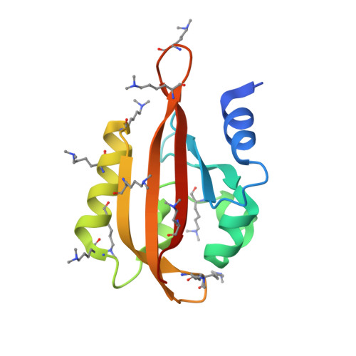
Structural properties of PAS domains from the KCNH potassium channels

Adaixo, R.Harley, C.A.Castro-Rodrigues, A.F.Morais-Cabral, J.H.

(2013) PLoS One 8: e59265-e59265

  • DOI: https://doi.org/10.1371/journal.pone.0059265
  • Primary Citation Related Structures: 
    4HOI, 4HP4, 4HP9, 4HQA

  • PubMed Abstract: 

    KCNH channels form an important family of voltage gated potassium channels. These channels include a N-terminal Per-Arnt-Sim (PAS) domain with unknown function. In other proteins PAS domains are implicated in cellular responses to environmental queues through small molecule binding or involvement in signaling cascades. To better understand their role we characterized the structural properties of several channel PAS domains. We determined high resolution structures of PAS domains from the mouse EAG (mEAG), drosophila ELK (dELK) and human ERG (hERG) channels and also of the hERG domain without the first nine amino acids. We analyzed these structures for features connected to ligand binding and signaling in other PAS domains. In particular, we have found cavities in the hERG and mEAG structures that share similarities with the ligand binding sites from other PAS domains. These cavities are lined by polar and apolar chemical groups and display potential flexibility in their volume. We have also found that the hydrophobic patch on the domain β-sheet is a conserved feature and appears to drive the formation of protein-protein contacts. In addition, the structures of the dELK domain and of the truncated hERG domain revealed the presence of N-terminal helices. These helices are equivalent to the helix described in the hERG NMR structures and are known to be important for channel function. Overall, these channel domains retain many of the PAS domain characteristics known to be important for cell signaling.


  • Organizational Affiliation
    • IBMC, Instituto de Biologia Molecular e Celular, Universidade do Porto, Porto, Portugal.

Macromolecule Content 

  • Total Structure Weight: 30.52 kDa 
  • Atom Count: 2,284 
  • Modeled Residue Count: 249 
  • Deposited Residue Count: 260 
  • Unique protein chains: 1

Macromolecules

Find similar proteins by:|  3D Structure
Entity ID: 1
MoleculeChains  Sequence LengthOrganismDetailsImage
Eag-like K[+] channel
A, B
130Drosophila melanogasterMutation(s): 0 
Gene Names: Dmel_CG5076elk
EC: 2.7.3
UniProt
Find proteins for A1ZB14 (Drosophila melanogaster)
Explore A1ZB14 
Go to UniProtKB:  A1ZB14
Entity Groups
Sequence Clusters30% Identity50% Identity70% Identity90% Identity95% Identity100% Identity
UniProt GroupA1ZB14
Sequence Annotations
Expand
Reference Sequence

Experimental Data & Validation

Experimental Data

  • Method: X-RAY DIFFRACTION
  • Resolution: 2.00 Å
  • R-Value Free:  0.191 (Depositor), 0.203 (DCC) 
  • R-Value Work:  0.173 (Depositor), 0.189 (DCC) 
  • R-Value Observed: 0.174 (Depositor) 
Space Group: P 41 3 2
Unit Cell:
Length ( Å )Angle ( ˚ )
a = 150.306α = 90
b = 150.306β = 90
c = 150.306γ = 90
Software Package:
Software NamePurpose
MOSFLMdata reduction
SCALAdata scaling
PHASERphasing
REFMACrefinement
PDB_EXTRACTdata extraction
PHENIXrefinement

Structure Validation

View Full Validation Report


Entry History 

Deposition Data

Revision History  (Full details and data files)

  • Version 1.0: 2013-03-27
    Type: Initial release
  • Version 1.1: 2013-06-19
    Changes: Database references
  • Version 1.2: 2023-11-08
    Changes: Data collection, Database references, Derived calculations, Refinement description