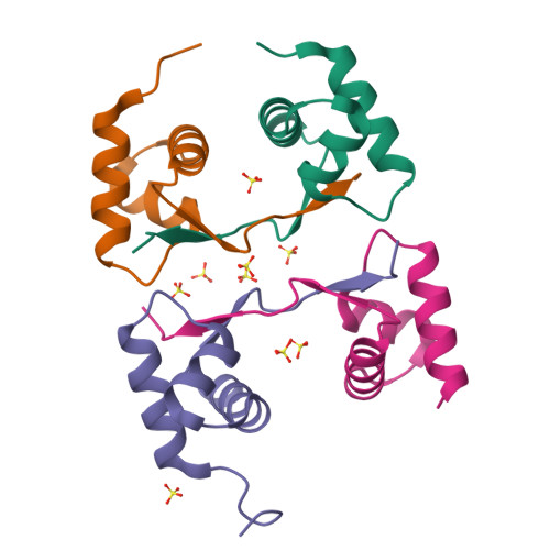
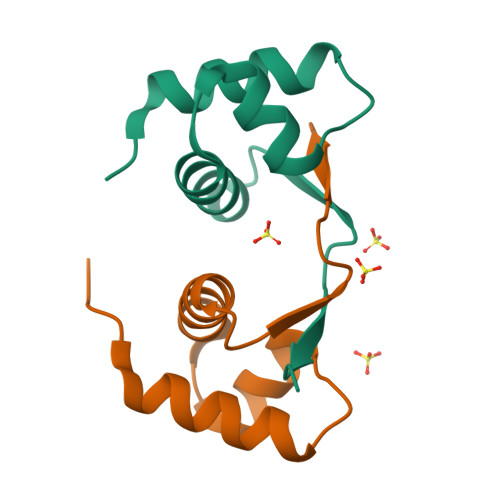
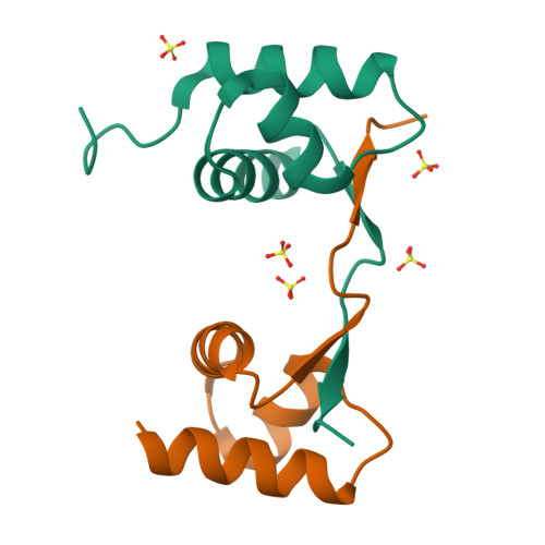
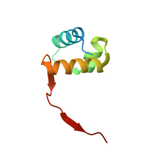
4HOB

The crystal structure of the Zalpha domain from Cyprinid Herpes virus 3


Experimental Data Snapshot

  • Method: X-RAY DIFFRACTION
  • Resolution: 1.76 Å
  • R-Value Free: 0.247 
  • R-Value Work: 0.201 
  • R-Value Observed: 0.204 

Starting Model: experimental
View more details

wwPDB Validation   3D Report Full Report


This is version 1.4 of the entry. See complete history


Literature

Crystal structure of a poxvirus-like zalpha domain from cyprinid herpesvirus 3

Tome, A.R.Kus, K.Correia, S.Paulo, L.M.Zacarias, S.de Rosa, M.Figueiredo, D.Parkhouse, R.M.Athanasiadis, A.

(2013) J Virol 87: 3998-4004

  • DOI: https://doi.org/10.1128/JVI.03116-12
  • Primary Citation of Related Structures:  
    4HOB

  • PubMed Abstract: 

    Zalpha domains are a subfamily of the winged helix-turn-helix domains sharing the unique ability to recognize CpG repeats in the left-handed Z-DNA conformation. In vertebrates, domains of this family are found exclusively in proteins that detect foreign nucleic acids and activate components of the antiviral interferon response. Moreover, poxviruses encode the Zalpha domain-containing protein E3L, a well-studied and potent inhibitor of interferon response. Here we describe a herpesvirus Zalpha-domain-containing protein (ORF112) from cyprinid herpesvirus 3. We demonstrate that ORF112 also binds CpG repeats in the left-handed conformation, and moreover, its structure at 1.75 Å reveals the Zalpha fold found in ADAR1, DAI, PKZ, and E3L. Unlike other Zalpha domains, however, ORF112 forms a dimer through a unique domain-swapping mechanism. Thus, ORF112 may be considered a new member of the Z-domain family having DNA binding properties similar to those of the poxvirus E3L inhibitor of interferon response.


  • Organizational Affiliation

    Instituto Gulbenkian de Ciência, Oeiras, Portugal.


Macromolecules
Find similar proteins by:  (by identity cutoff)  |  3D Structure
Entity ID: 1
MoleculeChains Sequence LengthOrganismDetailsImage
Putative uncharacterized protein
A, B, C, D
69Cyprinid herpesvirus 3Mutation(s): 0 
Gene Names: KHVJ122ORF112
UniProt
Find proteins for A4FTK7 (Cyprinid herpesvirus 3)
Explore A4FTK7 
Go to UniProtKB:  A4FTK7
Entity Groups  
Sequence Clusters30% Identity50% Identity70% Identity90% Identity95% Identity100% Identity
UniProt GroupA4FTK7
Sequence Annotations
Expand
  • Reference Sequence
Small Molecules
Ligands 1 Unique
IDChains Name / Formula / InChI Key2D Diagram3D Interactions
SO4
Query on SO4

Download Ideal Coordinates CCD File 
E [auth A]
F [auth A]
G [auth B]
H [auth B]
I [auth C]
E [auth A],
F [auth A],
G [auth B],
H [auth B],
I [auth C],
J [auth C],
K [auth C],
L [auth C],
M [auth C]
SULFATE ION
O4 S
QAOWNCQODCNURD-UHFFFAOYSA-L
Experimental Data & Validation

Experimental Data

  • Method: X-RAY DIFFRACTION
  • Resolution: 1.76 Å
  • R-Value Free: 0.247 
  • R-Value Work: 0.201 
  • R-Value Observed: 0.204 
  • Space Group: P 21 21 21
Unit Cell:
Length ( Å )Angle ( ˚ )
a = 51.736α = 90
b = 54.584β = 90
c = 86.642γ = 90
Software Package:
Software NamePurpose
DNAdata collection
PHASERphasing
PHENIXrefinement
XDSdata reduction
SCALAdata scaling

Structure Validation

View Full Validation Report


Entry History 

Deposition Data

Revision History  (Full details and data files)

  • Version 1.0: 2013-09-11
    Type: Initial release
  • Version 1.1: 2015-03-04
    Changes: Other
  • Version 1.2: 2019-12-04
    Changes: Data collection, Database references
  • Version 1.3: 2019-12-18
    Changes: Data collection
  • Version 1.4: 2023-11-08
    Changes: Data collection, Database references, Derived calculations, Refinement description