4HLU | pdb_00004hlu

Structure of the EcfA-A' heterodimer bound to ADP


Experimental Data Snapshot

  • Method: X-RAY DIFFRACTION
  • Resolution: 2.70 Å
  • R-Value Free: 
    0.258 (Depositor), 0.264 (DCC) 
  • R-Value Work: 
    0.213 (Depositor), 0.227 (DCC) 
  • R-Value Observed: 
    0.215 (Depositor) 

Starting Model: experimental
View more details

wwPDB Validation 3D Report Full Report

Validation slider image for 4HLU

Ligand Structure Quality Assessment 


This is version 1.4 of the entry. See complete history

Literature

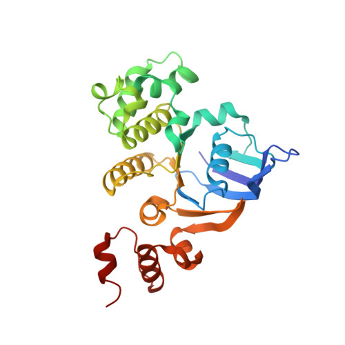
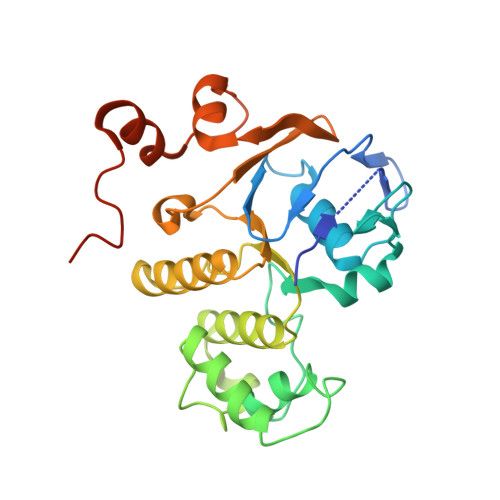
Assembly and mechanism of a group II ECF transporter.

Karpowich, N.K.Wang, D.N.

(2013) Proc Natl Acad Sci U S A 110: 2534-2539

  • DOI: https://doi.org/10.1073/pnas.1217361110
  • Primary Citation Related Structures: 
    4HLU

  • PubMed Abstract: 

    Energy-coupling factor (ECF) transporters are a recently discovered family of primary active transporters for micronutrients and vitamins, such as biotin, thiamine, and riboflavin. Found exclusively in archaea and bacteria, including the human pathogens Listeria, Streptococcus, and Staphylococcus, ECF transporters may be the only means of vitamin acquisition in these organisms. The subunit composition of ECF transporters is similar to that of ATP binding cassette (ABC) importers, whereby both systems share two homologous ATPase subunits (A and A'), a high affinity substrate-binding subunit (S), and a transmembrane coupling subunit (T). However, the S subunit of ECF transporters is an integral membrane protein, and the transmembrane coupling subunits do not share an obvious sequence homology between the two transporter families. Moreover, the subunit stoichiometry of ECF transporters is controversial, and the detailed molecular interactions between subunits and the conformational changes during substrate translocation are unknown. We have characterized the ECF transporters from Thermotoga maritima and Streptococcus thermophilus. Our data suggests a subunit stoichiometry of 2S:2T:1A:1A' and that S subunits for different substrates can be incorporated into the same transporter complex simultaneously. In the first crystal structure of the A-A' heterodimer, each subunit contains a novel motif called the Q-helix that plays a key role in subunit coupling with the T subunits. Taken together, these findings suggest a mechanism for coupling ATP binding and hydrolysis to transmembrane transport by ECF transporters.


  • Organizational Affiliation
    • The Helen L and Martin S Kimmel Center for Biology and Medicine, Skirball Institute of Biomolecular Medicine, and Department of Cell Biology, New York University School of Medicine, New York, NY 10016, USA. nathan.karpowich@med.nyu.edu

Macromolecule Content 

  • Total Structure Weight: 122.5 kDa 
  • Atom Count: 8,336 
  • Modeled Residue Count: 1,024 
  • Deposited Residue Count: 1,072 
  • Unique protein chains: 2

Macromolecules

Find similar proteins by:|  3D Structure
Entity ID: 1
MoleculeChains  Sequence LengthOrganismDetailsImage
Putative ABC transporter ATP-binding protein TM_0222A,
C [auth B]
268Thermotoga maritima MSB8Mutation(s): 0 
Gene Names: TM0222TM_0222
EC: 3.6.3 (PDB Primary Data), 7 (UniProt)
UniProt
Find proteins for Q9WY65 (Thermotoga maritima (strain ATCC 43589 / DSM 3109 / JCM 10099 / NBRC 100826 / MSB8))
Explore Q9WY65 
Go to UniProtKB:  Q9WY65
Entity Groups
Sequence Clusters30% Identity50% Identity70% Identity90% Identity95% Identity100% Identity
UniProt GroupQ9WY65
Sequence Annotations
Expand
Reference Sequence
Find similar proteins by:|  3D Structure
Entity ID: 2
MoleculeChains  Sequence LengthOrganismDetailsImage
Energy-coupling factor transporter ATP-binding protein EcfAB [auth D],
D [auth C]
268Thermotoga maritima MSB8Mutation(s): 7 
Gene Names: cbiOecfATM1663TM_1663
EC: 3.6.3 (PDB Primary Data), 7 (UniProt)
UniProt
Find proteins for Q9X1Z1 (Thermotoga maritima (strain ATCC 43589 / DSM 3109 / JCM 10099 / NBRC 100826 / MSB8))
Explore Q9X1Z1 
Go to UniProtKB:  Q9X1Z1
Entity Groups
Sequence Clusters30% Identity50% Identity70% Identity90% Identity95% Identity100% Identity
UniProt GroupQ9X1Z1
Sequence Annotations
Expand
Reference Sequence

Small Molecules

Ligands 2 Unique
IDChains Name / Formula / InChI Key2D Diagram3D Interactions
ADP

Query on ADP


Download:Ideal Coordinates CCD File
E [auth A],
G [auth D],
I [auth B],
K [auth C]
ADENOSINE-5'-DIPHOSPHATE
C10 H15 N5 O10 P2
XTWYTFMLZFPYCI-KQYNXXCUSA-N
ACT

Query on ACT


Download:Ideal Coordinates CCD File
F [auth A],
H [auth D],
J [auth B],
L [auth C],
M [auth C]
ACETATE ION
C2 H3 O2
QTBSBXVTEAMEQO-UHFFFAOYSA-M

Experimental Data & Validation

Experimental Data

  • Method: X-RAY DIFFRACTION
  • Resolution: 2.70 Å
  • R-Value Free:  0.258 (Depositor), 0.264 (DCC) 
  • R-Value Work:  0.213 (Depositor), 0.227 (DCC) 
  • R-Value Observed: 0.215 (Depositor) 
Space Group: P 32
Unit Cell:
Length ( Å )Angle ( ˚ )
a = 67.785α = 90
b = 67.785β = 90
c = 252.509γ = 120
Software Package:
Software NamePurpose
HKL-2000data collection
PHASERphasing
PHENIXrefinement
HKL-2000data reduction
HKL-2000data scaling

Structure Validation

View Full Validation Report


Ligand Structure Quality Assessment 


Entry History 

Deposition Data

Revision History  (Full details and data files)

  • Version 1.0: 2013-02-06
    Type: Initial release
  • Version 1.1: 2013-02-20
    Changes: Database references
  • Version 1.2: 2013-02-27
    Changes: Database references
  • Version 1.3: 2023-09-20
    Changes: Data collection, Database references, Derived calculations, Refinement description
  • Version 1.4: 2024-10-16
    Changes: Structure summary