4HH4 | pdb_00004hh4

Structure of the CcbJ Methyltransferase from Streptomyces caelestis


Experimental Data Snapshot

  • Method: X-RAY DIFFRACTION
  • Resolution: 2.90 Å
  • R-Value Free: 
    0.221 (Depositor), 0.214 (DCC) 
  • R-Value Work: 
    0.183 (Depositor), 0.176 (DCC) 
  • R-Value Observed: 
    0.185 (Depositor) 

wwPDB Validation 3D Report Full Report

Validation slider image for 4HH4

Ligand Structure Quality Assessment 


This is version 1.3 of the entry. See complete history

Literature

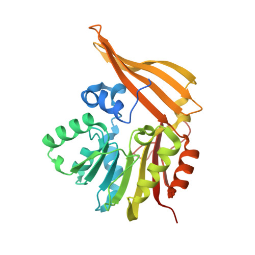
Structure and possible mechanism of the CcbJ methyltransferase from Streptomyces caelestis.

Bauer, J.Ondrovicova, G.Najmanova, L.Pevala, V.Kamenik, Z.Kostan, J.Janata, J.Kutejova, E.

(2014) Acta Crystallogr D Biol Crystallogr 70: 943-957

  • DOI: https://doi.org/10.1107/S139900471303397X
  • Primary Citation Related Structures: 
    4HGY, 4HGZ, 4HH4

  • PubMed Abstract: 

    The S-adenosyl-L-methionine (SAM)-dependent methyltransferase CcbJ from Streptomyces caelestis catalyzes one of the final steps in the biosynthesis of the antibiotic celesticetin, methylation of the N atom of its proline moiety, which greatly enhances the activity of the antibiotic. Since several celesticetin variants exist, this enzyme may be able to act on a variety of substrates. The structures of CcbJ determined by MAD phasing at 3.0 Å resolution, its native form at 2.7 Å resolution and its complex with S-adenosyl-L-homocysteine (SAH) at 2.9 Å resolution are reported here. Based on these structures, three point mutants, Y9F, Y17F and F117G, were prepared in order to study its behaviour as well as docking simulations of both CcbJ-SAM-substrate and CcbJ-SAH-product complexes. The structures show that CcbJ is a class I SAM-dependent methyltransferase with a wide active site, thereby suggesting that it may accommodate a number of different substrates. The mutation results show that the Y9F and F117G mutants are almost non-functional, while the Y17F mutant has almost half of the wild-type activity. In combination with the docking studies, these results suggest that Tyr9 and Phe117 are likely to help to position the substrate for the methyl-transfer reaction and that Tyr9 may also facilitate the reaction by removing an H(+) ion. Tyr17, on the other hand, seems to operate by helping to stabilize the SAM cofactor.


  • Organizational Affiliation
    • Institute of Molecular Biology, Slovak Academy of Sciences, 851 45 Bratislava, Slovakia.

Macromolecule Content 

  • Total Structure Weight: 185.33 kDa 
  • Atom Count: 12,077 
  • Modeled Residue Count: 1,519 
  • Deposited Residue Count: 1,656 
  • Unique protein chains: 1

Macromolecules

Find similar proteins by:|  3D Structure
Entity ID: 1
MoleculeChains  Sequence LengthOrganismDetailsImage
CcbJ
A, B, C, D, E
A, B, C, D, E, F
276Streptomyces caelestisMutation(s): 0 
Gene Names: ccbJ
UniProt
Find proteins for E9JES0 (Streptomyces caelestis)
Explore E9JES0 
Go to UniProtKB:  E9JES0
Entity Groups
Sequence Clusters30% Identity50% Identity70% Identity90% Identity95% Identity100% Identity
UniProt GroupE9JES0
Sequence Annotations
Expand
Reference Sequence

Small Molecules

Ligands 4 Unique
IDChains Name / Formula / InChI Key2D Diagram3D Interactions
SAH

Query on SAH


Download:Ideal Coordinates CCD File
BA [auth E]
G [auth A]
JA [auth F]
M [auth B]
R [auth C]
BA [auth E],
G [auth A],
JA [auth F],
M [auth B],
R [auth C],
X [auth D]
S-ADENOSYL-L-HOMOCYSTEINE
C14 H20 N6 O5 S
ZJUKTBDSGOFHSH-WFMPWKQPSA-N
EPE

Query on EPE


Download:Ideal Coordinates CCD File
Q [auth B]4-(2-HYDROXYETHYL)-1-PIPERAZINE ETHANESULFONIC ACID
C8 H18 N2 O4 S
JKMHFZQWWAIEOD-UHFFFAOYSA-N
SO4

Query on SO4


Download:Ideal Coordinates CCD File
CA [auth E]
DA [auth E]
EA [auth E]
FA [auth E]
GA [auth E]
CA [auth E],
DA [auth E],
EA [auth E],
FA [auth E],
GA [auth E],
H [auth A],
HA [auth E],
I [auth A],
J [auth A],
KA [auth F],
LA [auth F],
MA [auth F],
N [auth B],
O [auth B],
S [auth C],
T [auth C],
U [auth C],
V [auth C],
Y [auth D],
Z [auth D]
SULFATE ION
O4 S
QAOWNCQODCNURD-UHFFFAOYSA-L
GOL

Query on GOL


Download:Ideal Coordinates CCD File
AA [auth D]
IA [auth E]
K [auth A]
L [auth A]
P [auth B]
AA [auth D],
IA [auth E],
K [auth A],
L [auth A],
P [auth B],
W [auth C]
GLYCEROL
C3 H8 O3
PEDCQBHIVMGVHV-UHFFFAOYSA-N

Experimental Data & Validation

Experimental Data

  • Method: X-RAY DIFFRACTION
  • Resolution: 2.90 Å
  • R-Value Free:  0.221 (Depositor), 0.214 (DCC) 
  • R-Value Work:  0.183 (Depositor), 0.176 (DCC) 
  • R-Value Observed: 0.185 (Depositor) 
Space Group: C 2 2 21
Unit Cell:
Length ( Å )Angle ( ˚ )
a = 169.95α = 90
b = 244β = 90
c = 118.07γ = 90
Software Package:
Software NamePurpose
ADSCdata collection
PHASERphasing
REFMACrefinement
MOSFLMdata reduction
SCALAdata scaling

Structure Validation

View Full Validation Report


Ligand Structure Quality Assessment 


Entry History 

Deposition Data

Revision History  (Full details and data files)

  • Version 1.0: 2013-10-30
    Type: Initial release
  • Version 1.1: 2014-04-16
    Changes: Database references
  • Version 1.2: 2020-02-26
    Changes: Data collection, Database references
  • Version 1.3: 2024-02-28
    Changes: Data collection, Database references, Derived calculations