4HF0 | pdb_00004hf0

Crystal Structure of Apo IscR


Experimental Data Snapshot

  • Method: X-RAY DIFFRACTION
  • Resolution: 1.90 Å
  • R-Value Free: 
    0.221 (Depositor), 0.225 (DCC) 
  • R-Value Work: 
    0.193 (Depositor), 0.197 (DCC) 
  • R-Value Observed: 
    0.196 (Depositor) 

wwPDB Validation 3D Report Full Report

Validation slider image for 4HF0

This is version 1.3 of the entry. See complete history

Literature

Studies of IscR reveal a unique mechanism for metal-dependent regulation of DNA binding specificity.

Rajagopalan, S.Teter, S.J.Zwart, P.H.Brennan, R.G.Phillips, K.J.Kiley, P.J.

(2013) Nat Struct Mol Biol 20: 740-747

  • DOI: https://doi.org/10.1038/nsmb.2568
  • Primary Citation Related Structures: 
    4HF0, 4HF1, 4HF2

  • PubMed Abstract: 

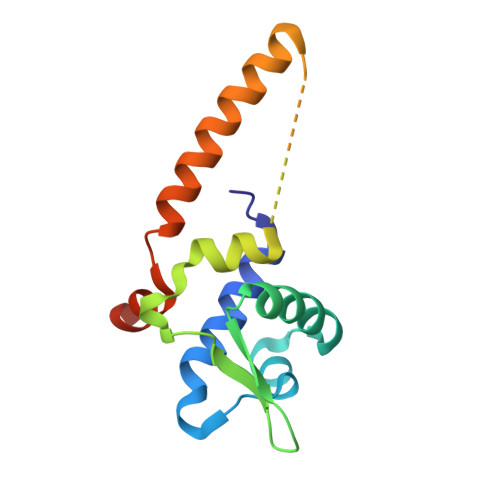
    IscR from Escherichia coli is an unusual metalloregulator in that both apo and iron sulfur (Fe-S)-IscR regulate transcription and exhibit different DNA binding specificities. Here, we report structural and biochemical studies of IscR suggesting that remodeling of the protein-DNA interface upon Fe-S ligation broadens the DNA binding specificity of IscR from binding the type 2 motif only to both type 1 and type 2 motifs. Analysis of an apo-IscR variant with relaxed target-site discrimination identified a key residue in wild-type apo-IscR that, we propose, makes unfavorable interactions with a type 1 motif. Upon Fe-S binding, these interactions are apparently removed, thereby allowing holo-IscR to bind both type 1 and type 2 motifs. These data suggest a unique mechanism of ligand-mediated DNA site recognition, whereby metallocluster ligation relocates a protein-specificity determinant to expand DNA target-site selection, allowing a broader transcriptomic response by holo-IscR.


  • Organizational Affiliation
    • Genomic Medicine Program, The Methodist Hospital Research Institute, Houston, Texas, USA.

Macromolecule Content 

  • Total Structure Weight: 29.96 kDa 
  • Atom Count: 1,785 
  • Modeled Residue Count: 238 
  • Deposited Residue Count: 282 
  • Unique protein chains: 1

Macromolecules

Find similar proteins by:|  3D Structure
Entity ID: 1
MoleculeChains  Sequence LengthOrganismDetailsImage
HTH-type transcriptional regulator IscR
A, B
141Escherichia coli K-12Mutation(s): 3 
Gene Names: iscRyfhPb2531JW2515
UniProt
Find proteins for P0AGK8 (Escherichia coli (strain K12))
Explore P0AGK8 
Go to UniProtKB:  P0AGK8
Entity Groups
Sequence Clusters30% Identity50% Identity70% Identity90% Identity95% Identity100% Identity
UniProt GroupP0AGK8
Sequence Annotations
Expand
Reference Sequence

Small Molecules

Ligands 1 Unique
IDChains Name / Formula / InChI Key2D Diagram3D Interactions
SO4

Query on SO4


Download:Ideal Coordinates CCD File
C [auth B]SULFATE ION
O4 S
QAOWNCQODCNURD-UHFFFAOYSA-L

Experimental Data & Validation

Experimental Data

  • Method: X-RAY DIFFRACTION
  • Resolution: 1.90 Å
  • R-Value Free:  0.221 (Depositor), 0.225 (DCC) 
  • R-Value Work:  0.193 (Depositor), 0.197 (DCC) 
  • R-Value Observed: 0.196 (Depositor) 
Space Group: P 31 2 1
Unit Cell:
Length ( Å )Angle ( ˚ )
a = 88.85α = 90
b = 88.85β = 90
c = 62.734γ = 120
Software Package:
Software NamePurpose
PHENIXrefinement
PDB_EXTRACTdata extraction
HKL-2000data reduction
SCALEPACKdata scaling
PHASERphasing

Structure Validation

View Full Validation Report


Entry History 

Deposition Data

Revision History  (Full details and data files)

  • Version 1.0: 2013-05-08
    Type: Initial release
  • Version 1.1: 2013-05-29
    Changes: Database references
  • Version 1.2: 2013-07-03
    Changes: Database references
  • Version 1.3: 2024-02-28
    Changes: Data collection, Database references, Derived calculations