4HE7 | pdb_00004he7

Crystal Structure of Brazzein


Experimental Data Snapshot

  • Method: X-RAY DIFFRACTION
  • Resolution: 1.80 Å
  • R-Value Free: 
    0.252 (Depositor), 0.250 (DCC) 
  • R-Value Work: 
    0.218 (Depositor), 0.225 (DCC) 
  • R-Value Observed: 
    0.219 (Depositor) 

Starting Model: experimental
View more details

wwPDB Validation 3D Report Full Report

Validation slider image for 4HE7

This is version 2.2 of the entry. See complete history

Literature

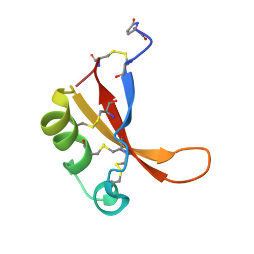
The structure of brazzein, a sweet-tasting protein from the wild African plant Pentadiplandra brazzeana

Nagata, K.Hongo, N.Kameda, Y.Yamamura, A.Sasaki, H.Lee, W.C.Ishikawa, K.Suzuki, E.Tanokura, M.

(2013) Acta Crystallogr D Biol Crystallogr 69: 642-647

  • DOI: https://doi.org/10.1107/S0907444913001005
  • Primary Citation Related Structures: 
    4HE7

  • PubMed Abstract: 

    Brazzein is the smallest sweet-tasting protein and was isolated from the wild African plant Pentadiplandra brazzeana. The brazzein molecule consists of 54 amino-acid residues and four disulfide bonds. Here, the first crystal structure of brazzein is reported at 1.8 Å resolution and is compared with previously reported solution structures. Despite the overall structural similarity, there are several remarkable differences between the crystal and solution structures both in their backbone folds and side-chain conformations. Firstly, there is an additional α-helix in the crystal structure. Secondly, the atomic r.m.s.d.s between the corresponding C(α)-atom pairs are as large as 2.0-2.2 Å between the crystal and solution structures. Thirdly, the crystal structure exhibits a molecular shape that is similar but not identical to the solution structures. The crystal structure of brazzein reported here will provide additional information and further insights into the intermolecular interaction of brazzein with the sweet-taste receptor.


  • Organizational Affiliation
    • Department of Applied Biological Chemistry, Graduate School of Agricultural and Life Sciences, The University of Tokyo, Tokyo, Japan.

Macromolecule Content 

  • Total Structure Weight: 6.51 kDa 
  • Atom Count: 463 
  • Modeled Residue Count: 54 
  • Deposited Residue Count: 54 
  • Unique protein chains: 1

Macromolecules

Find similar proteins by:|  3D Structure
Entity ID: 1
MoleculeChains  Sequence LengthOrganismDetailsImage
Defensin-like protein54Pentadiplandra brazzeanaMutation(s): 0 
UniProt
Find proteins for P56552 (Pentadiplandra brazzeana)
Explore P56552 
Go to UniProtKB:  P56552
Entity Groups
Sequence Clusters30% Identity50% Identity70% Identity90% Identity95% Identity100% Identity
UniProt GroupP56552
Sequence Annotations
Expand
Reference Sequence

Small Molecules

Ligands 1 Unique
IDChains Name / Formula / InChI Key2D Diagram3D Interactions
NA

Query on NA


Download:Ideal Coordinates CCD File
B [auth A]SODIUM ION
Na
FKNQFGJONOIPTF-UHFFFAOYSA-N
Modified Residues  1 Unique
IDChains TypeFormula2D DiagramParent
PCA
Query on PCA
A
L-PEPTIDE LINKINGC5 H7 N O3GLN

Experimental Data & Validation

Experimental Data

  • Method: X-RAY DIFFRACTION
  • Resolution: 1.80 Å
  • R-Value Free:  0.252 (Depositor), 0.250 (DCC) 
  • R-Value Work:  0.218 (Depositor), 0.225 (DCC) 
  • R-Value Observed: 0.219 (Depositor) 
Space Group: I 41 2 2
Unit Cell:
Length ( Å )Angle ( ˚ )
a = 61.4α = 90
b = 61.4β = 90
c = 59.6γ = 90
Software Package:
Software NamePurpose
WEISdata scaling
MOLREPphasing
REFMACrefinement
HKL-2000data reduction
HKL-2000data scaling

Structure Validation

View Full Validation Report


Entry History 

Deposition Data

Revision History  (Full details and data files)

  • Version 1.0: 2013-03-27
    Type: Initial release
  • Version 1.1: 2013-08-07
    Changes: Database references
  • Version 1.2: 2017-11-15
    Changes: Refinement description
  • Version 1.3: 2018-07-18
    Changes: Data collection, Database references
  • Version 2.0: 2019-12-25
    Changes: Database references, Derived calculations, Polymer sequence
  • Version 2.1: 2023-11-08
    Changes: Data collection, Database references, Derived calculations, Refinement description
  • Version 2.2: 2024-11-20
    Changes: Structure summary