4HD4 | pdb_00004hd4

Crystal Structure of Tyrosinase from Bacillus megaterium V218F mutant


Experimental Data Snapshot

  • Method: X-RAY DIFFRACTION
  • Resolution: 1.80 Å
  • R-Value Free: 
    0.228 (Depositor), 0.225 (DCC) 
  • R-Value Work: 
    0.207 (Depositor), 0.202 (DCC) 
  • R-Value Observed: 
    0.209 (Depositor) 

wwPDB Validation 3D Report Full Report

Validation slider image for 4HD4

This is version 1.2 of the entry. See complete history

Literature

Influencing the monophenolase/diphenolase activity ratio in tyrosinase.

Goldfeder, M.Kanteev, M.Adir, N.Fishman, A.

(2013) Biochim Biophys Acta 1834: 629-633

  • DOI: https://doi.org/10.1016/j.bbapap.2012.12.021
  • Primary Citation Related Structures: 
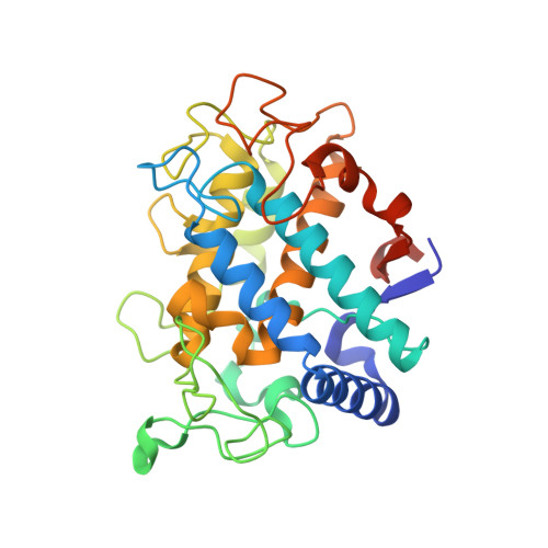
    4HD4, 4HD6, 4HD7

  • PubMed Abstract: 

    Tyrosinase is a type 3 copper enzyme with great potential for production of commercially valuable diphenols from monophenols. However, the use of tyrosinase is limited by its further oxidation of diphenols to quinones. We recently determined the structure of the Bacillus megaterium tyrosinase revealing a residue, V218, which we proposed to take part in positioning of substrates within the active site. In the structure of catechol oxidase from Ipomoea batatas, the lack of monophenolase activity was attributed to the presence of F261 near CuA. Consequently, we engineered two variants, V218F and V218G. V218F was expected to have a decreased monophenolase activity, due to the bulky residue extending into the active site. Surprisingly, both V218F and V218G exhibited a 9- and 4.4-fold higher monophenolase/diphenolase activity ratio, respectively. X-ray structures of variant V218F display a flexibility of the phenylalanine residue along with an adjacent histidine, which we propose to be the source of the change in activity ratio.


  • Organizational Affiliation
    • Department of Biotechnology and Food Engineering, Technion-Israel Institute of Technology, Haifa 32000, Israel.

Macromolecule Content 

  • Total Structure Weight: 70.73 kDa 
  • Atom Count: 4,956 
  • Modeled Residue Count: 573 
  • Deposited Residue Count: 606 
  • Unique protein chains: 1

Macromolecules

Find similar proteins by:|  3D Structure
Entity ID: 1
MoleculeChains  Sequence LengthOrganismDetailsImage
Tyrosinase
A, B
303Priestia megateriumMutation(s): 2 
UniProt
Find proteins for B2ZB02 (Priestia megaterium)
Explore B2ZB02 
Go to UniProtKB:  B2ZB02
Entity Groups
Sequence Clusters30% Identity50% Identity70% Identity90% Identity95% Identity100% Identity
UniProt GroupB2ZB02
Sequence Annotations
Expand
Reference Sequence

Experimental Data & Validation

Experimental Data

  • Method: X-RAY DIFFRACTION
  • Resolution: 1.80 Å
  • R-Value Free:  0.228 (Depositor), 0.225 (DCC) 
  • R-Value Work:  0.207 (Depositor), 0.202 (DCC) 
  • R-Value Observed: 0.209 (Depositor) 
Space Group: P 1 21 1
Unit Cell:
Length ( Å )Angle ( ˚ )
a = 48α = 90
b = 78.58β = 106.13
c = 85.89γ = 90
Software Package:
Software NamePurpose
MOSFLMdata reduction
SCALAdata scaling
PHASERphasing
PHENIXrefinement
PDB_EXTRACTdata extraction

Structure Validation

View Full Validation Report


Entry History 

Deposition Data

Revision History  (Full details and data files)

  • Version 1.0: 2013-01-23
    Type: Initial release
  • Version 1.1: 2013-02-13
    Changes: Database references
  • Version 1.2: 2024-02-28
    Changes: Data collection, Database references, Derived calculations