The Structure of Classical Swine Fever Virus N(pro): A Novel Cysteine Autoprotease and Zinc-Binding Protein Involved in Subversion of Type I Interferon Induction.
Gottipati, K., Ruggli, N., Gerber, M., Tratschin, J.D., Benning, M., Bellamy, H., Choi, K.H.(2013) PLoS Pathog 9: e1003704-e1003704
- PubMed: 24146623 Search on PubMedSearch on PubMed Central
- DOI: https://doi.org/10.1371/journal.ppat.1003704
- Primary Citation Related Structures:
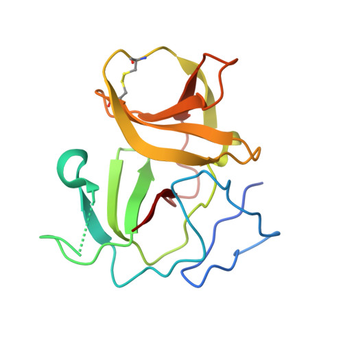
4H9J, 4H9K - PubMed Abstract:
Pestiviruses express their genome as a single polypeptide that is subsequently cleaved into individual proteins by host- and virus-encoded proteases. The pestivirus N-terminal protease (N(pro)) is a cysteine autoprotease that cleaves between its own C-terminus and the N-terminus of the core protein. Due to its unique sequence and catalytic site, it forms its own cysteine protease family C53. After self-cleavage, N(pro) is no longer active as a protease. The released N(pro) suppresses the induction of the host's type-I interferon-α/β (IFN-α/β) response. N(pro) binds interferon regulatory factor-3 (IRF3), the key transcriptional activator of IFN-α/β genes, and promotes degradation of IRF3 by the proteasome, thus preventing induction of the IFN-α/β response to pestivirus infection. Here we report the crystal structures of pestivirus N(pro). N(pro) is structurally distinct from other known cysteine proteases and has a novel "clam shell" fold consisting of a protease domain and a zinc-binding domain. The unique fold of N(pro) allows auto-catalysis at its C-terminus and subsequently conceals the cleavage site in the active site of the protease. Although many viruses interfere with type I IFN induction by targeting the IRF3 pathway, little information is available regarding structure or mechanism of action of viral proteins that interact with IRF3. The distribution of amino acids on the surface of N(pro) involved in targeting IRF3 for proteasomal degradation provides insight into the nature of N(pro)'s interaction with IRF3. The structures thus establish the mechanism of auto-catalysis and subsequent auto-inhibition of trans-activity of N(pro), and its role in subversion of host immune response.
- Department of Biochemistry and Molecular Biology, Sealy Center for Structural Biology and Molecular Biophysics, The University of Texas Medical Branch at Galveston, Galveston, Texas, United States of America.
Organizational Affiliation: