


TBD
Chi, B., Zaccai, N.R., Brady, R.L., Woolfson, D.N.To be published.
Experimental Data Snapshot
Starting Model: experimental
View more details
wwPDB Validation 3D Report Full Report

Entity ID: 1 | |||||
|---|---|---|---|---|---|
| Molecule | Chains | Sequence Length | Organism | Details | Image |
| CC-Hex-H24-A5/7C | 32 | synthetic construct | Mutation(s): 0 | 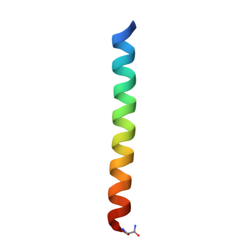 | |
| Length ( Å ) | Angle ( ˚ ) |
|---|---|
| a = 31.7 | α = 90 |
| b = 31.7 | β = 90 |
| c = 132.04 | γ = 120 |
| Software Name | Purpose |
|---|---|
| PHENIX | refinement |


