4H8G | pdb_00004h8g

crystal structure of a parallel 6-helix coiled coil CC-Hex-IL- 22


Experimental Data Snapshot

  • Method: X-RAY DIFFRACTION
  • Resolution: 1.70 Å
  • R-Value Free: 
    0.280 (Depositor), 0.274 (DCC) 
  • R-Value Work: 
    0.247 (Depositor), 0.241 (DCC) 
  • R-Value Observed: 
    0.249 (Depositor) 

wwPDB Validation 3D Report Full Report

Validation slider image for 4H8G

This is version 1.1 of the entry. See complete history

Literature

TBA

Chi, B.Zaccai, N.R.Brady, R.L.Woolfson, D.N.

(null) J Am Chem Soc 

Macromolecule Content 

  • Total Structure Weight: 39.86 kDa 
  • Atom Count: 2,985 
  • Modeled Residue Count: 359 
  • Deposited Residue Count: 384 
  • Unique protein chains: 1

Macromolecules

Find similar proteins by:|  3D Structure
Entity ID: 1
MoleculeChains  Sequence LengthOrganismDetailsImage
CC-Hex-IL-2232synthetic constructMutation(s): 0 

Small Molecules

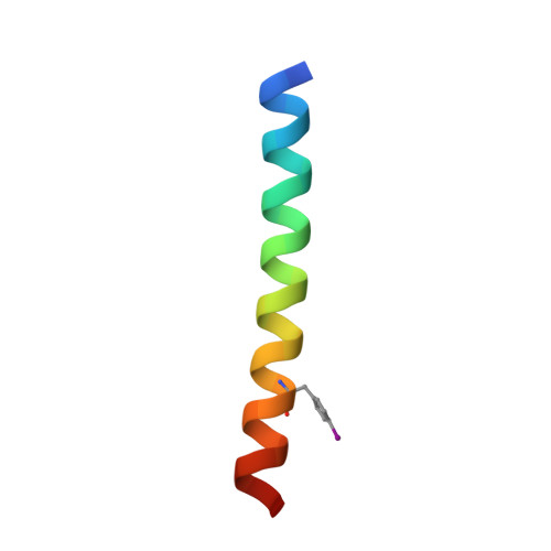
Modified Residues  1 Unique
IDChains TypeFormula2D DiagramParent
PHI
Query on PHI
A
B
C
D
E
A,
B,
C,
D,
E,
F,
G [auth I],
H [auth J],
I [auth G],
J [auth H],
K,
L
L-PEPTIDE LINKINGC9 H10 I N O2PHE

Experimental Data & Validation

Experimental Data

  • Method: X-RAY DIFFRACTION
  • Resolution: 1.70 Å
  • R-Value Free:  0.280 (Depositor), 0.274 (DCC) 
  • R-Value Work:  0.247 (Depositor), 0.241 (DCC) 
  • R-Value Observed: 0.249 (Depositor) 
Space Group: C 2 2 21
Unit Cell:
Length ( Å )Angle ( ˚ )
a = 51.99α = 90
b = 126.8β = 90
c = 127.53γ = 90
Software Package:
Software NamePurpose
REFMACrefinement

Structure Validation

View Full Validation Report


Entry History 

Deposition Data

Revision History  (Full details and data files)

  • Version 1.0: 2014-02-05
    Type: Initial release
  • Version 1.1: 2025-03-26
    Changes: Data collection, Database references, Derived calculations, Structure summary