4H5X

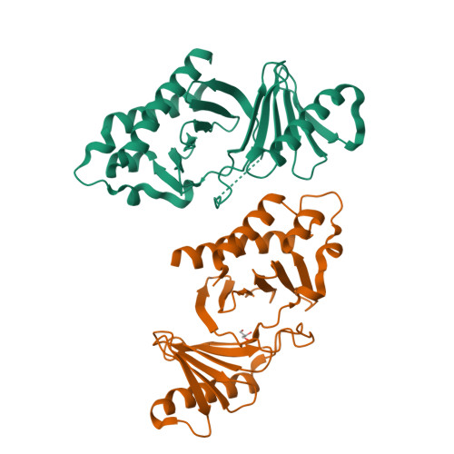
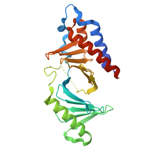
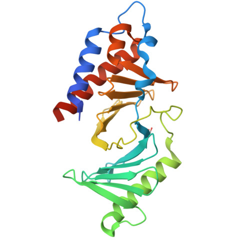
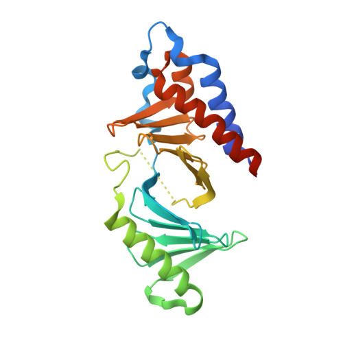
human Plk1-PBD with a glycerol bound at the phophopeptide binding site


Experimental Data Snapshot

  • Method: X-RAY DIFFRACTION
  • Resolution: 1.95 Å
  • R-Value Free: 0.242 
  • R-Value Work: 0.198 
  • R-Value Observed: 0.200 

Starting Model: experimental
View more details

wwPDB Validation   3D Report Full Report


This is version 1.2 of the entry. See complete history


Literature

Thymoquinone Blocks pSer/pThr Recognition by Plk1 Polo-Box Domain As a Phosphate Mimic

Yin, Z.Song, Y.Rehse, P.H.

(2013) ACS Chem Biol 8: 303-308

  • DOI: https://doi.org/10.1021/cb3004379
  • Primary Citation of Related Structures:  
    4H5X, 4H71, 4HCO

  • PubMed Abstract: 

    Phosphorylation-dependent protein-protein interaction has rarely been targeted in medicinal chemistry. Thymoquinone, a naturally occurring antitumor agent, disrupts prephosphorylated substrate recognition by the polo-box domain of polo-like kinase 1, a key mitotic regulator responsible for various carcinogenesis when overexpressed. Here, crystallographic studies reveal that the phosphoserine/phosphothreonine recognition site of the polo-box domain is the binding pocket for thymoquinone and its analogue poloxime. Both small molecules displace phosphopeptides bound with the polo-box domain in a slow but noncovalent binding mode. A conserved water bridge and a cation-π interaction were found as their competition strategy against the phosphate group. This mechanism sheds light on small-molecule intervention of phospho-recognition by the polo-box domain of polo-like kinase 1 and other phospho-binding proteins in general.


  • Organizational Affiliation

    School of Life Science and Technology, China Pharmaceutical University, 24 Tong Jia Xiang, Nanjing, Jiangsu 210009, China.


Macromolecules
Find similar proteins by:  (by identity cutoff)  |  3D Structure
Entity ID: 1
MoleculeChains Sequence LengthOrganismDetailsImage
Serine/threonine-protein kinase PLK1A [auth B],
B [auth A]
240Homo sapiensMutation(s): 0 
Gene Names: PLK1PLK
EC: 2.7.11.21
UniProt & NIH Common Fund Data Resources
Find proteins for P53350 (Homo sapiens)
Explore P53350 
Go to UniProtKB:  P53350
PHAROS:  P53350
GTEx:  ENSG00000166851 
Entity Groups  
Sequence Clusters30% Identity50% Identity70% Identity90% Identity95% Identity100% Identity
UniProt GroupP53350
Sequence Annotations
Expand
  • Reference Sequence
Small Molecules
Ligands 1 Unique
IDChains Name / Formula / InChI Key2D Diagram3D Interactions
GOL
Query on GOL

Download Ideal Coordinates CCD File 
C [auth A]GLYCEROL
C3 H8 O3
PEDCQBHIVMGVHV-UHFFFAOYSA-N
Experimental Data & Validation

Experimental Data

  • Method: X-RAY DIFFRACTION
  • Resolution: 1.95 Å
  • R-Value Free: 0.242 
  • R-Value Work: 0.198 
  • R-Value Observed: 0.200 
  • Space Group: P 1 21 1
Unit Cell:
Length ( Å )Angle ( ˚ )
a = 33.3α = 90
b = 102.54β = 94.11
c = 67.58γ = 90
Software Package:
Software NamePurpose
REFMACrefinement
MOSFLMdata reduction
SCALAdata scaling

Structure Validation

View Full Validation Report


Entry History 

Deposition Data

Revision History  (Full details and data files)

  • Version 1.0: 2012-10-03
    Type: Initial release
  • Version 1.1: 2013-03-06
    Changes: Database references
  • Version 1.2: 2023-11-08
    Changes: Data collection, Database references, Derived calculations, Refinement description