4H3S | pdb_00004h3s

The Structure of Glutaminyl-tRNA Synthetase from Saccharomyces Cerevisiae


Experimental Data Snapshot

  • Method: X-RAY DIFFRACTION
  • Resolution: 2.15 Å
  • R-Value Free: 
    0.176 (Depositor), 0.180 (DCC) 
  • R-Value Work: 
    0.160 (Depositor), 0.165 (DCC) 
  • R-Value Observed: 
    0.161 (Depositor) 

Starting Model: experimental
View more details

wwPDB Validation 3D Report Full Report

Validation slider image for 4H3S

This is version 1.4 of the entry. See complete history

Literature

The Structure of Yeast Glutaminyl-tRNA Synthetase and Modeling of Its Interaction with tRNA.

Grant, T.D.Luft, J.R.Wolfley, J.R.Snell, M.E.Tsuruta, H.Corretore, S.Quartley, E.Phizicky, E.M.Grayhack, E.J.Snell, E.H.

(2013) J Mol Biology 425: 2480-2493

  • DOI: https://doi.org/10.1016/j.jmb.2013.03.043
  • Primary Citation Related Structures: 
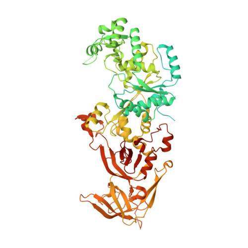
    4H3S

  • PubMed Abstract: 

    Eukaryotic glutaminyl-tRNA synthetase (GlnRS) contains an appended N-terminal domain (NTD) whose precise function is unknown. Although GlnRS structures from two prokaryotic species are known, no eukaryotic GlnRS structure has been reported. Here we present the first crystallographic structure of yeast GlnRS, finding that the structure of the C-terminal domain is highly similar to Escherichia coli GlnRS but that 214 residues, including the NTD, are crystallographically disordered. We present a model of the full-length enzyme in solution, using the structures of the C-terminal domain, and the isolated NTD, with small-angle X-ray scattering data of the full-length molecule. We proceed to model the enzyme bound to tRNA, using the crystallographic structures of GatCAB and GlnRS-tRNA complex from bacteria. We contrast the tRNA-bound model with the tRNA-free solution state and perform molecular dynamics on the full-length GlnRS-tRNA complex, which suggests that tRNA binding involves the motion of a conserved hinge in the NTD.


  • Organizational Affiliation
    • Hauptman Woodward Medical Research Institute, 700 Ellicott Street, Buffalo, NY 14203, USA.

Macromolecule Content 

  • Total Structure Weight: 95.43 kDa 
  • Atom Count: 5,553 
  • Modeled Residue Count: 585 
  • Deposited Residue Count: 817 
  • Unique protein chains: 1

Macromolecules

Find similar proteins by:|  3D Structure
Entity ID: 1
MoleculeChains  Sequence LengthOrganismDetailsImage
Glutamine-tRNA ligase817Saccharomyces cerevisiae S288CMutation(s): 0 
Gene Names: GLN4O3601YOR168W
EC: 6.1.1.18
UniProt
Find proteins for P13188 (Saccharomyces cerevisiae (strain ATCC 204508 / S288c))
Explore P13188 
Go to UniProtKB:  P13188
Entity Groups
Sequence Clusters30% Identity50% Identity70% Identity90% Identity95% Identity100% Identity
UniProt GroupP13188
Sequence Annotations
Expand
Reference Sequence

Small Molecules

Ligands 6 Unique
IDChains Name / Formula / InChI Key2D Diagram3D Interactions
DTT

Query on DTT


Download:Ideal Coordinates CCD File
Q [auth A]2,3-DIHYDROXY-1,4-DITHIOBUTANE
C4 H10 O2 S2
VHJLVAABSRFDPM-IMJSIDKUSA-N
GOL

Query on GOL


Download:Ideal Coordinates CCD File
C [auth A]
D [auth A]
E [auth A]
F [auth A]
G [auth A]
C [auth A],
D [auth A],
E [auth A],
F [auth A],
G [auth A],
H [auth A],
I [auth A],
J [auth A]
GLYCEROL
C3 H8 O3
PEDCQBHIVMGVHV-UHFFFAOYSA-N
BR

Query on BR


Download:Ideal Coordinates CCD File
K [auth A]BROMIDE ION
Br
CPELXLSAUQHCOX-UHFFFAOYSA-M
ZN

Query on ZN


Download:Ideal Coordinates CCD File
B [auth A]ZINC ION
Zn
PTFCDOFLOPIGGS-UHFFFAOYSA-N
ACT

Query on ACT


Download:Ideal Coordinates CCD File
M [auth A],
N [auth A],
O [auth A],
P [auth A]
ACETATE ION
C2 H3 O2
QTBSBXVTEAMEQO-UHFFFAOYSA-M
CL

Query on CL


Download:Ideal Coordinates CCD File
L [auth A]CHLORIDE ION
Cl
VEXZGXHMUGYJMC-UHFFFAOYSA-M

Experimental Data & Validation

Experimental Data

  • Method: X-RAY DIFFRACTION
  • Resolution: 2.15 Å
  • R-Value Free:  0.176 (Depositor), 0.180 (DCC) 
  • R-Value Work:  0.160 (Depositor), 0.165 (DCC) 
  • R-Value Observed: 0.161 (Depositor) 
Space Group: P 31 2 1
Unit Cell:
Length ( Å )Angle ( ˚ )
a = 176.611α = 90
b = 176.611β = 90
c = 72.188γ = 120
Software Package:
Software NamePurpose
Blu-Icedata collection
PHENIXmodel building
PHENIXrefinement
XDSdata reduction
SCALAdata scaling
PHENIXphasing

Structure Validation

View Full Validation Report


Entry History 

Deposition Data

Revision History  (Full details and data files)

  • Version 1.0: 2013-04-24
    Type: Initial release
  • Version 1.1: 2013-07-31
    Changes: Database references
  • Version 1.2: 2014-11-12
    Changes: Structure summary
  • Version 1.3: 2014-11-26
    Changes: Other
  • Version 1.4: 2023-09-20
    Changes: Data collection, Database references, Derived calculations, Refinement description