4H2X | pdb_00004h2x

Crystal structure of engineered Bradyrhizobium japonicum glycine:[carrier protein] ligase complexed with carrier protein from Agrobacterium tumefaciens and an analogue of glycyl adenylate


Experimental Data Snapshot

  • Method: X-RAY DIFFRACTION
  • Resolution: 2.15 Å
  • R-Value Free: 
    0.198 (Depositor), 0.199 (DCC) 
  • R-Value Work: 
    0.167 (Depositor), 0.171 (DCC) 
  • R-Value Observed: 
    0.168 (Depositor) 

wwPDB Validation 3D Report Full Report

Validation slider image for 4H2X

Ligand Structure Quality Assessment 


This is version 1.5 of the entry. See complete history

Literature

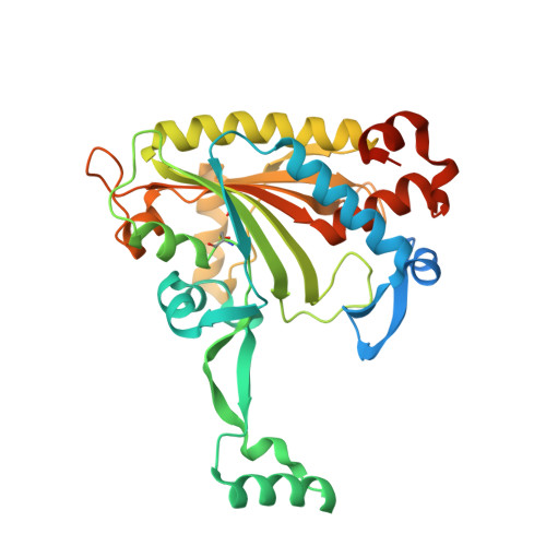
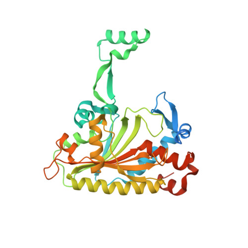
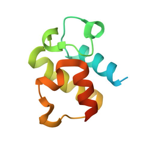
Adaptation of aminoacyl-tRNA synthetase catalytic core to carrier protein aminoacylation.

Mocibob, M.Ivic, N.Luic, M.Weygand-Durasevic, I.

(2013) Structure 21: 614-626

  • DOI: https://doi.org/10.1016/j.str.2013.02.017
  • Primary Citation Related Structures: 
    4H2S, 4H2T, 4H2U, 4H2V, 4H2W, 4H2X, 4H2Y

  • PubMed Abstract: 

    Amino acid:[carrier protein] ligases (aa:CP ligases) are recently discovered enzymes that are highly similar to class II aminoacyl-tRNA synthetases (aaRSs). However, while aaRSs aminoacylate tRNA and supply building blocks for ribosomal translation, aa:CP ligases transfer activated amino acids to the phosphopantetheine group of small carrier proteins. We have solved the crystal structure of an aa:CP ligase complexed with the carrier protein (CP). The CP prosthetic group enters the active site from a different direction than tRNA in class II aaRS complexes through an idiosyncratic tunnel. CP binds to aa:CP ligase in a fundamentally different manner compared to tRNA binding by structurally closely related aaRSs. Based on crystallographic analysis, an enzyme of altered CP specificity was designed, and the mechanism of amino acid transfer to the prosthetic group was proposed. The presented study reveals how a conserved class II aaRS catalytic core can adapt to another function through minor structural alterations.


  • Organizational Affiliation
    • Department of Chemistry, Faculty of Science, University of Zagreb, Horvatovac 102a, 10 000 Zagreb, Croatia.

Macromolecule Content 

  • Total Structure Weight: 100.37 kDa 
  • Atom Count: 6,243 
  • Modeled Residue Count: 743 
  • Deposited Residue Count: 898 
  • Unique protein chains: 3

Macromolecules

Find similar proteins by:|  3D Structure
Entity ID: 1
MoleculeChains  Sequence LengthOrganismDetailsImage
Amino acid--[acyl-carrier-protein] ligase 1346Bradyrhizobium diazoefficiens USDA 110Agrobacterium fabrum str. C58Mutation(s): 0 
Gene Names: bll0957Atu2573AGR_C_4663
EC: 6.2.1
UniProt
Find proteins for Q89VT8 (Bradyrhizobium diazoefficiens (strain JCM 10833 / BCRC 13528 / IAM 13628 / NBRC 14792 / USDA 110))
Explore Q89VT8 
Go to UniProtKB:  Q89VT8
Entity Groups
Sequence Clusters30% Identity50% Identity70% Identity90% Identity95% Identity100% Identity
UniProt GroupQ89VT8
Sequence Annotations
Expand
Reference Sequence
Find similar proteins by:|  3D Structure
Entity ID: 2
MoleculeChains  Sequence LengthOrganismDetailsImage
Amino acid--[acyl-carrier-protein] ligase 1346Bradyrhizobium diazoefficiens USDA 110Agrobacterium fabrum str. C58Mutation(s): 0 
Gene Names: bll0957Atu2573AGR_C_4663
EC: 6.2.1
UniProt
Find proteins for Q89VT8 (Bradyrhizobium diazoefficiens (strain JCM 10833 / BCRC 13528 / IAM 13628 / NBRC 14792 / USDA 110))
Explore Q89VT8 
Go to UniProtKB:  Q89VT8
Entity Groups
Sequence Clusters30% Identity50% Identity70% Identity90% Identity95% Identity100% Identity
UniProt GroupQ89VT8
Sequence Annotations
Expand
Reference Sequence
Find similar proteins by:|  3D Structure
Entity ID: 3
MoleculeChains  Sequence LengthOrganismDetailsImage
Aminoacyl carrier protein
C, D
103Agrobacterium fabrum str. C58Mutation(s): 0 
Gene Names: AGR_C_4658Atu2571
UniProt
Find proteins for A9CHM9 (Agrobacterium fabrum (strain C58 / ATCC 33970))
Explore A9CHM9 
Go to UniProtKB:  A9CHM9
Entity Groups
Sequence Clusters30% Identity50% Identity70% Identity90% Identity95% Identity100% Identity
UniProt GroupA9CHM9
Sequence Annotations
Expand
Reference Sequence

Small Molecules

Ligands 4 Unique
IDChains Name / Formula / InChI Key2D Diagram3D Interactions
G5A

Query on G5A


Download:Ideal Coordinates CCD File
E [auth A],
H [auth B]
5'-O-(glycylsulfamoyl)adenosine
C12 H17 N7 O7 S
AMWPZASLDLLQFT-JJNLEZRASA-N
PNS

Query on PNS


Download:Ideal Coordinates CCD File
J [auth C],
K [auth D]
4'-PHOSPHOPANTETHEINE
C11 H23 N2 O7 P S
JDMUPRLRUUMCTL-VIFPVBQESA-N
ZN

Query on ZN


Download:Ideal Coordinates CCD File
G [auth B]ZINC ION
Zn
PTFCDOFLOPIGGS-UHFFFAOYSA-N
CL

Query on CL


Download:Ideal Coordinates CCD File
F [auth A],
I [auth B]
CHLORIDE ION
Cl
VEXZGXHMUGYJMC-UHFFFAOYSA-M
Modified Residues  1 Unique
IDChains TypeFormula2D DiagramParent
CSO
Query on CSO
A
L-PEPTIDE LINKINGC3 H7 N O3 SCYS

Experimental Data & Validation

Experimental Data

  • Method: X-RAY DIFFRACTION
  • Resolution: 2.15 Å
  • R-Value Free:  0.198 (Depositor), 0.199 (DCC) 
  • R-Value Work:  0.167 (Depositor), 0.171 (DCC) 
  • R-Value Observed: 0.168 (Depositor) 
Space Group: P 21 21 21
Unit Cell:
Length ( Å )Angle ( ˚ )
a = 99.575α = 90
b = 101.433β = 90
c = 103.044γ = 90
Software Package:
Software NamePurpose
XSCALEdata scaling
PHENIXrefinement
PDB_EXTRACTdata extraction
DNAdata collection
XDSdata reduction
PHENIXphasing

Structure Validation

View Full Validation Report


Ligand Structure Quality Assessment 


Entry History 

Deposition Data

Revision History  (Full details and data files)

  • Version 1.0: 2013-03-06
    Type: Initial release
  • Version 1.1: 2013-04-10
    Changes: Database references
  • Version 1.2: 2013-05-29
    Changes: Database references
  • Version 1.3: 2013-06-19
    Changes: Atomic model
  • Version 1.4: 2017-08-23
    Changes: Refinement description, Source and taxonomy
  • Version 1.5: 2025-03-26
    Changes: Data collection, Database references, Derived calculations, Structure summary