4H1J | pdb_00004h1j

Crystal structure of PYK2 with the pyrazole 13a


Experimental Data Snapshot

  • Method: X-RAY DIFFRACTION
  • Resolution: 2.00 Å
  • R-Value Free: 
    0.209 (Depositor), 0.220 (DCC) 
  • R-Value Work: 
    0.180 (Depositor), 0.180 (DCC) 
  • R-Value Observed: 
    0.182 (Depositor) 

wwPDB Validation   3D Report Full Report


Ligand Structure Quality Assessment 

Created with Raphaël 2.3.0Worse 01 BetterLigand structure goodness of fit to experimental dataBest fitted 0YHClick on this verticalbar to view details

This is version 1.2 of the entry. See complete history


Literature

Identification of novel series of pyrazole and indole-urea based DFG-out PYK2 inhibitors.

Bhattacharya, S.K.Aspnes, G.E.Bagley, S.W.Boehm, M.Brosius, A.D.Buckbinder, L.Chang, J.S.Dibrino, J.Eng, H.Frederick, K.S.Griffith, D.A.Griffor, M.C.Guimaraes, C.R.Guzman-Perez, A.Han, S.Kalgutkar, A.S.Klug-McLeod, J.Garcia-Irizarry, C.Li, J.Lippa, B.Price, D.A.Southers, J.A.Walker, D.P.Wei, L.Xiao, J.Zawistoski, M.P.Zhao, X.

(2012) Bioorg Med Chem Lett 22: 7523-7529

  • DOI: https://doi.org/10.1016/j.bmcl.2012.10.039
  • Primary Citation of Related Structures:  
    4H1J, 4H1M

  • PubMed Abstract: 

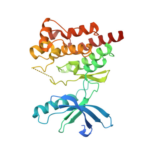
    Previous drug discovery efforts identified classical PYK2 kinase inhibitors such as 2 and 3 that possess selectivity for PYK2 over its intra-family isoform FAK. Efforts to identify more kinome-selective chemical matter that stabilize a DFG-out conformation of the enzyme are described herein. Two sub-series of PYK2 inhibitors, an indole carboxamide-urea and a pyrazole-urea have been identified and found to have different binding interactions with the hinge region of PYK2. These leads proved to be more selective than the original classical inhibitors.


  • Organizational Affiliation

    Worldwide Medicinal Chemistry, Pfizer Global Research and Development, 620 Memorial Drive, Cambridge, MA 02139, United States. samit.k.bhattacharya@pfizer.com


Macromolecules
Find similar proteins by:  (by identity cutoff)  |  3D Structure
Entity ID: 1
MoleculeChains Sequence LengthOrganismDetailsImage
Protein-tyrosine kinase 2-beta293Homo sapiensMutation(s): 0 
Gene Names: PTK2BFAK2PYK2RAFTK
EC: 2.7.10.2
UniProt & NIH Common Fund Data Resources
Find proteins for Q14289 (Homo sapiens)
Explore Q14289 
Go to UniProtKB:  Q14289
PHAROS:  Q14289
GTEx:  ENSG00000120899 
Entity Groups  
Sequence Clusters30% Identity50% Identity70% Identity90% Identity95% Identity100% Identity
UniProt GroupQ14289
Sequence Annotations
Expand
  • Reference Sequence
Small Molecules
Ligands 1 Unique
IDChains Name / Formula / InChI Key2D Diagram3D Interactions
0YH
Query on 0YH

Download Ideal Coordinates CCD File 
B [auth A]1-[3-tert-butyl-1-(4-methylphenyl)-1H-pyrazol-5-yl]-3-[3-(4-methoxy-2-methylphenyl)-1H-pyrazol-5-yl]urea
C26 H30 N6 O2
LATBWIWKSPOPNK-UHFFFAOYSA-N
Binding Affinity Annotations 
IDSourceBinding Affinity
0YH BindingDB:  4H1J IC50: 1200 (nM) from 1 assay(s)
Experimental Data & Validation

Experimental Data

  • Method: X-RAY DIFFRACTION
  • Resolution: 2.00 Å
  • R-Value Free:  0.209 (Depositor), 0.220 (DCC) 
  • R-Value Work:  0.180 (Depositor), 0.180 (DCC) 
  • R-Value Observed: 0.182 (Depositor) 
Space Group: P 21 21 21
Unit Cell:
Length ( Å )Angle ( ˚ )
a = 37.945α = 90
b = 82.923β = 90
c = 85.728γ = 90
Software Package:
Software NamePurpose
HKL-2000data collection
PHASERphasing
BUSTERrefinement
HKL-2000data reduction
SCALEPACKdata scaling

Structure Validation

View Full Validation Report


Ligand Structure Quality Assessment 

Created with Raphaël 2.3.0Worse 01 BetterLigand structure goodness of fit to experimental dataBest fitted 0YHClick on this verticalbar to view details

Entry History 

Deposition Data

  • Released Date: 2012-11-28 
  • Deposition Author(s): Han, S.

Revision History  (Full details and data files)

  • Version 1.0: 2012-11-28
    Type: Initial release
  • Version 1.1: 2012-12-12
    Changes: Database references
  • Version 1.2: 2024-02-28
    Changes: Data collection, Database references, Derived calculations