4H0P | pdb_00004h0p

Crystal Structure of Acetate Kinase from Cryptococcus neoformans


Experimental Data Snapshot

  • Method: X-RAY DIFFRACTION
  • Resolution: 1.89 Å
  • R-Value Free: 
    0.219 (Depositor), 0.225 (DCC) 
  • R-Value Work: 
    0.178 (Depositor), 0.184 (DCC) 
  • R-Value Observed: 
    0.180 (Depositor) 

Starting Model: experimental
View more details

wwPDB Validation 3D Report Full Report

Validation slider image for 4H0P

This is version 1.2 of the entry. See complete history

Literature

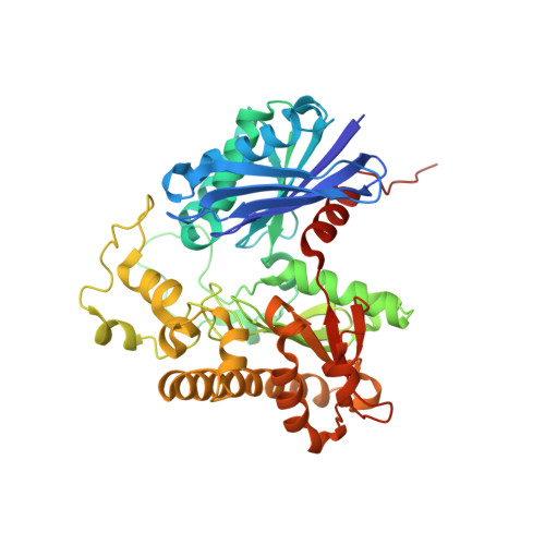
Crystal structures of acetate kinases from the eukaryotic pathogens Entamoeba histolytica and Cryptococcus neoformans.

Thaker, T.M.Tanabe, M.Fowler, M.L.Preininger, A.M.Ingram-Smith, C.Smith, K.S.Iverson, T.M.

(2013) J Struct Biol 181: 185-189

  • DOI: https://doi.org/10.1016/j.jsb.2012.11.001
  • Primary Citation Related Structures: 
    4H0O, 4H0P

  • PubMed Abstract: 

    Acetate kinases (ACKs) are members of the acetate and sugar kinase/hsp70/actin (ASKHA) superfamily and catalyze the reversible phosphorylation of acetate, with ADP/ATP the most common phosphoryl acceptor/donor. While prokaryotic ACKs have been the subject of extensive biochemical and structural characterization, there is a comparative paucity of information on eukaryotic ACKs, and prior to this report, no structure of an ACK of eukaryotic origin was available. We determined the structures of ACKs from the eukaryotic pathogens Entamoeba histolytica and Cryptococcus neoformans. Each active site is located at an interdomain interface, and the acetate and phosphate binding pockets display sequence and structural conservation with their prokaryotic counterparts. Interestingly, the E. histolytica ACK has previously been shown to be pyrophosphate (PP(i))-dependent, and is the first ACK demonstrated to have this property. Examination of its structure demonstrates how subtle amino acid substitutions within the active site have converted cosubstrate specificity from ATP to PP(i) while retaining a similar backbone conformation. Differences in the angle between domains surrounding the active site suggest that interdomain movement may accompany catalysis. Taken together, these structures are consistent with the eukaryotic ACKs following a similar reaction mechanism as is proposed for the prokaryotic homologs.


  • Organizational Affiliation
    • Department of Biochemistry, Vanderbilt University Medical Center, Nashville, TN 37232, USA.

Macromolecule Content 

  • Total Structure Weight: 96.35 kDa 
  • Atom Count: 7,034 
  • Modeled Residue Count: 861 
  • Deposited Residue Count: 876 
  • Unique protein chains: 1

Macromolecules

Find similar proteins by:|  3D Structure
Entity ID: 1
MoleculeChains  Sequence LengthOrganismDetailsImage
acetate kinase
A, B
438Cryptococcus neoformansMutation(s): 0 
Gene Names: ACKA
EC: 2.7.2.1
UniProt
Find proteins for J9W3A6 (Cryptococcus neoformans (strain H99 / ATCC 208821 / CBS 10515 / FGSC 9487))
Explore J9W3A6 
Go to UniProtKB:  J9W3A6
Entity Groups
Sequence Clusters30% Identity50% Identity70% Identity90% Identity95% Identity100% Identity
UniProt GroupJ9W3A6
Sequence Annotations
Expand
Reference Sequence

Experimental Data & Validation

Experimental Data

  • Method: X-RAY DIFFRACTION
  • Resolution: 1.89 Å
  • R-Value Free:  0.219 (Depositor), 0.225 (DCC) 
  • R-Value Work:  0.178 (Depositor), 0.184 (DCC) 
  • R-Value Observed: 0.180 (Depositor) 
Space Group: P 1 21 1
Unit Cell:
Length ( Å )Angle ( ˚ )
a = 51.37α = 90
b = 107.613β = 99.83
c = 79.108γ = 90
Software Package:
Software NamePurpose
HKL-2000data collection
PHASERphasing
PHENIXrefinement
HKL-2000data reduction
SCALEPACKdata scaling

Structure Validation

View Full Validation Report


Entry History 

Deposition Data

Revision History  (Full details and data files)

  • Version 1.0: 2012-12-05
    Type: Initial release
  • Version 1.1: 2013-02-20
    Changes: Database references
  • Version 1.2: 2023-09-13
    Changes: Data collection, Database references, Refinement description