4GYE | pdb_00004gye

MDR 769 HIV-1 Protease in Complex with Reduced P1F


Experimental Data Snapshot

  • Method: X-RAY DIFFRACTION
  • Resolution: 2.27 Å
  • R-Value Free: 
    0.264 (Depositor), 0.259 (DCC) 
  • R-Value Work: 
    0.193 (Depositor), 0.195 (DCC) 
  • R-Value Observed: 
    0.196 (Depositor) 

Starting Model: experimental
View more details

wwPDB Validation 3D Report Full Report

Validation slider image for 4GYE

This is version 3.1 of the entry. See complete history

Literature

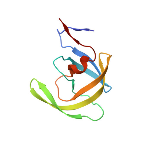
Ligand modifications to reduce the relative resistance of multi-drug resistant HIV-1 protease.

Dewdney, T.G.Wang, Y.Liu, Z.Sharma, S.K.Reiter, S.J.Brunzelle, J.S.Kovari, I.A.Woster, P.M.Kovari, L.C.

(2013) Bioorg Med Chem 21: 7430-7434

  • DOI: https://doi.org/10.1016/j.bmc.2013.09.045
  • Primary Citation Related Structures: 
    4GYE, 4GZF

  • PubMed Abstract: 

    Proper proteolytic processing of the HIV-1 Gag/Pol polyprotein is required for HIV infection and viral replication. This feature has made HIV-1 protease an attractive target for antiretroviral drug design for the treatment of HIV-1 infected patients. To examine the role of the P1 and P1'positions of the substrate in inhibitory efficacy of multi-drug resistant HIV-1 protease 769 (MDR 769), we performed a series of structure-function studies. Using the original CA/p2 cleavage site sequence, we generated heptapeptides containing one reduced peptide bond with an L to F and A to F double mutation at P1 and P1' (F-r-F), and an A to F at P1' (L-r-F) resulting in P1/P1' modified ligands. Here, we present an analysis of co-crystal structures of CA/p2 F-r-F, and CA/p2 L-r-F in complex with MDR 769. To examine conformational changes in the complex structure, molecular dynamic (MD) simulations were performed with MDR769-ligand complexes. MD trajectories show the isobutyl group of both the lopinavir analog and the CA/p2 L-r-F substrate cause a conformational change of in the active site of MDR 769. IC50 measurements suggest the non identical P1/P1' ligands (CA/p2 L-r-F and lopinavir analog) are more effective against MDR proteases as opposed to identical P1/P1'ligands. Our results suggest that a non identical P1/P1'composition may be more favorable for the inhibition of MDR 769 as they induce conformational changes in the active site of the enzyme resulting in disruption of the two-fold symmetry of the protease, thus, stabilizing the inhibitor in the active site.


  • Organizational Affiliation
    • Department of Biochemistry and Molecular Biology, School of Medicine, Wayne State University, Detroit, MI 48201, USA.

Macromolecule Content 

  • Total Structure Weight: 22.41 kDa 
  • Atom Count: 1,687 
  • Modeled Residue Count: 204 
  • Deposited Residue Count: 204 
  • Unique protein chains: 2

Macromolecules

Find similar proteins by:|  3D Structure
Entity ID: 1
MoleculeChains  Sequence LengthOrganismDetailsImage
Protease
A, B
99Human immunodeficiency virus 1Mutation(s): 5 
Gene Names: pol
UniProt
Find proteins for Q9QM22 (Human immunodeficiency virus type 1)
Explore Q9QM22 
Go to UniProtKB:  Q9QM22
Entity Groups
Sequence Clusters30% Identity50% Identity70% Identity90% Identity95% Identity100% Identity
UniProt GroupQ9QM22
Sequence Annotations
Expand
Reference Sequence
Find similar proteins by:  Sequence   |   3D Structure  
Entity ID: 2
MoleculeChains  Sequence LengthOrganismDetailsImage
P1F peptide6synthetic constructMutation(s): 0 

Small Molecules

Modified Residues  1 Unique
IDChains TypeFormula2D DiagramParent
NLE
Query on NLE
C
L-PEPTIDE LINKINGC6 H13 N O2LEU
PUK
Query on PUK
C
PEPTIDE-LIKEC18 H22 N2 O2

--

Experimental Data & Validation

Experimental Data

  • Method: X-RAY DIFFRACTION
  • Resolution: 2.27 Å
  • R-Value Free:  0.264 (Depositor), 0.259 (DCC) 
  • R-Value Work:  0.193 (Depositor), 0.195 (DCC) 
  • R-Value Observed: 0.196 (Depositor) 
Space Group: P 61
Unit Cell:
Length ( Å )Angle ( ˚ )
a = 62.255α = 90
b = 62.255β = 90
c = 83.92γ = 120
Software Package:
Software NamePurpose
HKL-2000data collection
REFMACrefinement

Structure Validation

View Full Validation Report


Entry History 

Deposition Data

Revision History  (Full details and data files)

  • Version 1.0: 2013-10-30
    Type: Initial release
  • Version 1.1: 2013-11-13
    Changes: Database references
  • Version 2.0: 2023-02-22
    Changes: Advisory, Atomic model, Data collection, Database references, Derived calculations, Non-polymer description, Polymer sequence, Source and taxonomy, Structure summary
  • Version 2.1: 2023-09-20
    Changes: Data collection, Refinement description
  • Version 3.0: 2023-11-15
    Changes: Atomic model, Data collection
  • Version 3.1: 2024-11-27
    Changes: Structure summary