4GXE | pdb_00004gxe

T. vulcanus Phycocyanin crystallized in 4M Urea


Experimental Data Snapshot

  • Method: X-RAY DIFFRACTION
  • Resolution: 3.00 Å
  • R-Value Free: 
    0.299 (Depositor), 0.244 (DCC) 
  • R-Value Work: 
    0.255 (Depositor), 0.246 (DCC) 
  • R-Value Observed: 
    0.255 (Depositor) 

Starting Model: experimental
View more details

wwPDB Validation 3D Report Full Report

Validation slider image for 4GXE

Ligand Structure Quality Assessment 


This is version 1.2 of the entry. See complete history

Literature

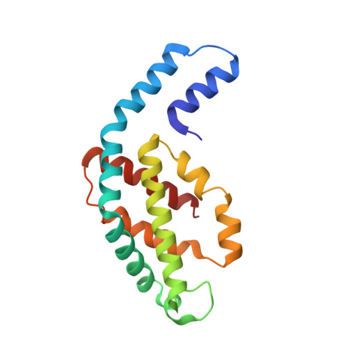
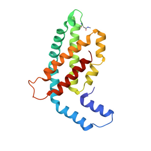
Allophycocyanin and phycocyanin crystal structures reveal facets of phycobilisome assembly.

Marx, A.Adir, N.

(2013) Biochim Biophys Acta 1827: 311-318

  • DOI: https://doi.org/10.1016/j.bbabio.2012.11.006
  • Primary Citation Related Structures: 
    4F0T, 4F0U, 4GXE, 4GY3, 4H0M

  • PubMed Abstract: 

    X-ray crystal structures of the isolated phycobiliprotein components of the phycobilisome have provided high resolution details to the description of this light harvesting complex at different levels of complexity and detail. The linker-independent assembly of trimers into hexamers in crystal lattices of previously determined structures has been observed in almost all of the phycocyanin (PC) and allophycocyanin (APC) structures available in the Protein Data Bank. In this paper we describe the X-ray crystal structures of PC and APC from Synechococcus elongatus sp. PCC 7942, PC from Synechocystis sp. PCC 6803 and PC from Thermosynechococcus vulcanus crystallized in the presence of urea. All five structures are highly similar to other PC and APC structures on the levels of subunits, monomers and trimers. The Synechococcus APC forms a unique loose hexamer that may show the structural requirements for core assembly and rod attachment. While the Synechococcus PC assembles into the canonical hexamer, it does not further assemble into rods. Unlike most PC structures, the Synechocystis PC fails to form hexamers. Addition of low concentrations of urea to T. vulcanus PC inhibits this proteins propensity to form hexamers, resulting in a crystal lattice composed of trimers. The molecular source of these differences in assembly and their relevance to the phycobilisome structure is discussed.


  • Organizational Affiliation
    • Schulich Faculty of Chemistry, Technion-Israel Institute of Technology, Haifa, Israel.

Macromolecule Content 

  • Total Structure Weight: 37.87 kDa 
  • Atom Count: 2,701 
  • Modeled Residue Count: 334 
  • Deposited Residue Count: 334 
  • Unique protein chains: 2

Macromolecules

Find similar proteins by:|  3D Structure
Entity ID: 1
MoleculeChains  Sequence LengthOrganismDetailsImage
C-phycocyanin alpha subunit162Thermostichus vulcanusMutation(s): 0 
UniProt
Find proteins for Q9AM02 (Thermostichus vulcanus)
Explore Q9AM02 
Go to UniProtKB:  Q9AM02
Entity Groups
Sequence Clusters30% Identity50% Identity70% Identity90% Identity95% Identity100% Identity
UniProt GroupQ9AM02
Sequence Annotations
Expand
Reference Sequence
Find similar proteins by:|  3D Structure
Entity ID: 2
MoleculeChains  Sequence LengthOrganismDetailsImage
C-phycocyanin beta subunit172Thermostichus vulcanusMutation(s): 0 
UniProt
Find proteins for Q71RW8 (Thermostichus vulcanus)
Explore Q71RW8 
Go to UniProtKB:  Q71RW8
Entity Groups
Sequence Clusters30% Identity50% Identity70% Identity90% Identity95% Identity100% Identity
UniProt GroupQ71RW8
Sequence Annotations
Expand
Reference Sequence

Small Molecules

Ligands 2 Unique
IDChains Name / Formula / InChI Key2D Diagram3D Interactions
CYC

Query on CYC


Download:Ideal Coordinates CCD File
C [auth A],
G [auth B],
H [auth B]
PHYCOCYANOBILIN
C33 H40 N4 O6
VXTXPYZGDQPMHK-GMXXPEQVSA-N
URE

Query on URE


Download:Ideal Coordinates CCD File
D [auth A]
E [auth A]
F [auth A]
I [auth B]
J [auth B]
D [auth A],
E [auth A],
F [auth A],
I [auth B],
J [auth B],
K [auth B],
L [auth B]
UREA
C H4 N2 O
XSQUKJJJFZCRTK-UHFFFAOYSA-N
Modified Residues  1 Unique
IDChains TypeFormula2D DiagramParent
MEN
Query on MEN
B
L-PEPTIDE LINKINGC5 H10 N2 O3ASN

Experimental Data & Validation

Experimental Data

  • Method: X-RAY DIFFRACTION
  • Resolution: 3.00 Å
  • R-Value Free:  0.299 (Depositor), 0.244 (DCC) 
  • R-Value Work:  0.255 (Depositor), 0.246 (DCC) 
  • R-Value Observed: 0.255 (Depositor) 
Space Group: P 63
Unit Cell:
Length ( Å )Angle ( ˚ )
a = 153.75α = 90
b = 153.75β = 90
c = 39.72γ = 120
Software Package:
Software NamePurpose
ADSCdata collection
PHASERphasing
CNSrefinement
MOSFLMdata reduction
SCALAdata scaling

Structure Validation

View Full Validation Report


Ligand Structure Quality Assessment 


Entry History 

Deposition Data

Revision History  (Full details and data files)

  • Version 1.0: 2013-03-06
    Type: Initial release
  • Version 1.1: 2013-03-27
    Changes: Other
  • Version 1.2: 2024-04-03
    Changes: Data collection, Database references, Derived calculations, Refinement description