4GWD | pdb_00004gwd

Crystal Structure of the Mn2+2,Zn2+-Human Arginase I-ABH Complex


Experimental Data Snapshot

  • Method: X-RAY DIFFRACTION
  • Resolution: 1.53 Å
  • R-Value Free: 
    0.215 (Depositor), 0.213 (DCC) 
  • R-Value Work: 
    0.160 (Depositor), 0.160 (DCC) 
  • R-Value Observed: 
    0.160 (Depositor) 

Starting Model: experimental
View more details

wwPDB Validation 3D Report Full Report

Validation slider image for 4GWD

Ligand Structure Quality Assessment 


This is version 1.2 of the entry. See complete history

Literature

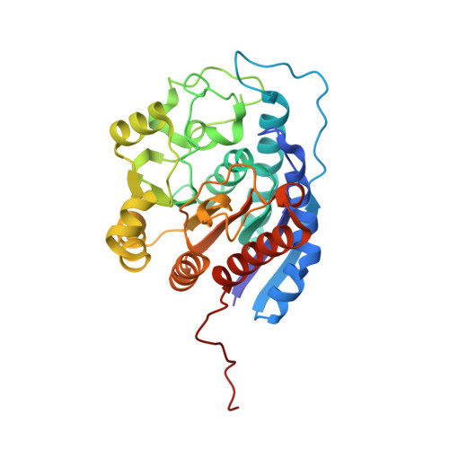
Structure and function of non-native metal clusters in human arginase I.

D'Antonio, E.L.Hai, Y.Christianson, D.W.

(2012) Biochemistry 51: 8399-8409

  • DOI: https://doi.org/10.1021/bi301145n
  • Primary Citation Related Structures: 
    4GSM, 4GSV, 4GSZ, 4GWC, 4GWD

  • PubMed Abstract: 

    Various binuclear metal ion clusters and complexes have been reconstituted in crystalline human arginase I by removing the Mn(2+)(2) cluster of the wild-type enzyme with metal chelators and subsequently soaking the crystalline apoenzyme in buffer solutions containing NiCl(2) or ZnCl(2). X-ray crystal structures of these metal ion variants are correlated with catalytic activity measurements that reveal differences resulting from metal ion substitution. Additionally, treatment of crystalline Mn(2+)(2)-human arginase I with Zn(2+) reveals for the first time the structural basis for inhibition by Zn(2+), which forms a carboxylate-histidine-Zn(2+) triad with H141 and E277. The imidazole side chain of H141 is known to be hyper-reactive, and its chemical modification or mutagenesis is known to similarly compromise catalysis. The reactive substrate analogue 2(S)-amino-6-boronohexanoic acid (ABH) binds as a tetrahedral boronate anion to Mn(2+)(2), Co(2+)(2), Ni(2+)(2), and Zn(2+)(2) clusters in human arginase I, and it can be stabilized by a third inhibitory Zn(2+) ion coordinated by H141. Because ABH binds as an analogue of the tetrahedral intermediate and its flanking transition states in catalysis, this implies that the various metallo-substituted enzymes are capable of some level of catalysis with an actual substrate. Accordingly, we establish the following trend for turnover number (k(cat)) and catalytic efficiency (k(cat)/K(M)): Mn(2+) > Ni(2+) ≈ Co(2+) ≫ Zn(2+). Therefore, Mn(2+) is required for optimal catalysis by human arginase I.


  • Organizational Affiliation
    • Roy and Diana Vagelos Laboratories, Department of Chemistry, University of Pennsylvania, Philadelphia, PA 19104-6323, USA.

Macromolecule Content 

  • Total Structure Weight: 70.29 kDa 
  • Atom Count: 4,965 
  • Modeled Residue Count: 626 
  • Deposited Residue Count: 644 
  • Unique protein chains: 1

Macromolecules

Find similar proteins by:|  3D Structure
Entity ID: 1
MoleculeChains  Sequence LengthOrganismDetailsImage
Arginase-1
A, B
322Homo sapiensMutation(s): 0 
Gene Names: ARG1
EC: 3.5.3.1
UniProt & NIH Common Fund Data Resources
Find proteins for P05089 (Homo sapiens)
Explore P05089 
Go to UniProtKB:  P05089
PHAROS:  P05089
GTEx:  ENSG00000118520 
Entity Groups
Sequence Clusters30% Identity50% Identity70% Identity90% Identity95% Identity100% Identity
UniProt GroupP05089
Sequence Annotations
Expand
Reference Sequence

Small Molecules

Ligands 3 Unique
IDChains Name / Formula / InChI Key2D Diagram3D Interactions
ABH

Query on ABH


Download:Ideal Coordinates CCD File
F [auth A],
J [auth B]
2(S)-AMINO-6-BORONOHEXANOIC ACID
C6 H15 B N O5
BLVGFZFOWWBCCZ-YFKPBYRVSA-N
ZN

Query on ZN


Download:Ideal Coordinates CCD File
E [auth A],
I [auth B]
ZINC ION
Zn
PTFCDOFLOPIGGS-UHFFFAOYSA-N
MN

Query on MN


Download:Ideal Coordinates CCD File
C [auth A],
D [auth A],
G [auth B],
H [auth B]
MANGANESE (II) ION
Mn
WAEMQWOKJMHJLA-UHFFFAOYSA-N

Experimental Data & Validation

Experimental Data

  • Method: X-RAY DIFFRACTION
  • Resolution: 1.53 Å
  • R-Value Free:  0.215 (Depositor), 0.213 (DCC) 
  • R-Value Work:  0.160 (Depositor), 0.160 (DCC) 
  • R-Value Observed: 0.160 (Depositor) 
Space Group: P 3
Unit Cell:
Length ( Å )Angle ( ˚ )
a = 91.082α = 90
b = 91.082β = 90
c = 69.903γ = 120
Software Package:
Software NamePurpose
CBASSdata collection
CNSrefinement
HKL-2000data reduction
HKL-2000data scaling
CNSphasing

Structure Validation

View Full Validation Report


Ligand Structure Quality Assessment 


Entry History 

Deposition Data

Revision History  (Full details and data files)

  • Version 1.0: 2012-09-26
    Type: Initial release
  • Version 1.1: 2012-11-14
    Changes: Database references
  • Version 1.2: 2023-09-13
    Changes: Data collection, Database references, Derived calculations, Refinement description