4GPM | pdb_00004gpm

Crystal Structure of Engineered Protein. Northeast Structural Genomics Consortium Target OR264.


Experimental Data Snapshot

  • Method: X-RAY DIFFRACTION
  • Resolution: 2.00 Å
  • R-Value Free: 
    0.202 (Depositor), 0.194 (DCC) 
  • R-Value Work: 
    0.176 (Depositor), 0.176 (DCC) 
  • R-Value Observed: 
    0.177 (Depositor) 

Starting Model: experimental
View more details

wwPDB Validation 3D Report Full Report

Validation slider image for 4GPM

This is version 1.3 of the entry. See complete history

Literature

Computational design of self-assembling cyclic protein homo-oligomers.

Fallas, J.A.Ueda, G.Sheffler, W.Nguyen, V.McNamara, D.E.Sankaran, B.Pereira, J.H.Parmeggiani, F.Brunette, T.J.Cascio, D.Yeates, T.R.Zwart, P.Baker, D.

(2017) Nat Chem 9: 353-360

  • DOI: https://doi.org/10.1038/nchem.2673
  • Primary Citation Related Structures: 
    4GMR, 4GPM, 4HB5, 4HXT, 5HRY, 5HRZ, 5HS0, 5K7V, 5KBA, 5KWD

  • PubMed Abstract: 

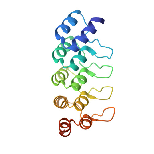
    Self-assembling cyclic protein homo-oligomers play important roles in biology, and the ability to generate custom homo-oligomeric structures could enable new approaches to probe biological function. Here we report a general approach to design cyclic homo-oligomers that employs a new residue-pair-transform method to assess the designability of a protein-protein interface. This method is sufficiently rapid to enable the systematic enumeration of cyclically docked arrangements of a monomer followed by sequence design of the newly formed interfaces. We use this method to design interfaces onto idealized repeat proteins that direct their assembly into complexes that possess cyclic symmetry. Of 96 designs that were characterized experimentally, 21 were found to form stable monodisperse homo-oligomers in solution, and 15 (four homodimers, six homotrimers, six homotetramers and one homopentamer) had solution small-angle X-ray scattering data consistent with the design models. X-ray crystal structures were obtained for five of the designs and each is very close to their corresponding computational model.


  • Organizational Affiliation
    • Department of Biochemistry, University of Washington, Seattle, Washington 98195, USA.

Macromolecule Content 

  • Total Structure Weight: 36.7 kDa 
  • Atom Count: 2,535 
  • Modeled Residue Count: 314 
  • Deposited Residue Count: 338 
  • Unique protein chains: 1

Macromolecules

Find similar proteins by:|  3D Structure
Entity ID: 1
MoleculeChains  Sequence LengthOrganismDetailsImage
Engineered Protein OR264
A, B
169synthetic constructMutation(s): 0 

Experimental Data & Validation

Experimental Data

  • Method: X-RAY DIFFRACTION
  • Resolution: 2.00 Å
  • R-Value Free:  0.202 (Depositor), 0.194 (DCC) 
  • R-Value Work:  0.176 (Depositor), 0.176 (DCC) 
  • R-Value Observed: 0.177 (Depositor) 
Space Group: P 21 21 21
Unit Cell:
Length ( Å )Angle ( ˚ )
a = 48.086α = 90
b = 56.824β = 90
c = 105.595γ = 90
Software Package:
Software NamePurpose
PHENIXrefinement
PDB_EXTRACTdata extraction
ADSCdata collection
HKL-2000data reduction
HKL-2000data scaling
BALBESphasing

Structure Validation

View Full Validation Report


Entry History 

Revision History  (Full details and data files)

  • Version 1.0: 2012-09-05
    Type: Initial release
  • Version 1.1: 2017-04-12
    Changes: Database references
  • Version 1.2: 2018-01-24
    Changes: Structure summary
  • Version 1.3: 2023-09-13
    Changes: Data collection, Database references, Refinement description