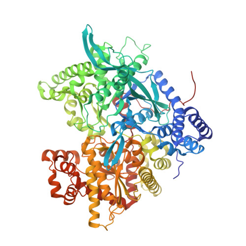
Comparison of the binding of glucose and glucose 1-phosphate derivatives to T-state glycogen phosphorylase b.
Martin, J.L., Johnson, L.N., Withers, S.G.(1990) Biochemistry 29: 10745-10757
- PubMed: 2125493
- DOI: https://doi.org/10.1021/bi00500a005
- Primary Citation of Related Structures:
2GPB, 3GPB, 4GPB, 5GPB - PubMed Abstract:
The binding of T-state- and R-state-stabilizing ligands to the catalytic C site of T-state glycogen phosphorylase b has been investigated by crystallographic methods to study the interactions made and the conformational changes that occur at the C site. The compounds studied were alpha-D-glucose, 1, a T-state-stabilizing inhibitor of the enzyme, and the R-state-stabilizing phosphorylated ligands alpha-D-glucose 1-phosphate (2), 2-deoxy-2-fluoro-alpha-D-glucose 1-phosphate (3), and alpha-D-glucose 1-methylenephosphonate (4). The complexes have been refined, giving crystallographic R factors of less than 19%, for data between 8 and 2.3 A. Analysis of the refined structures shows that the glucosyl portions of the phosphorylated ligands bind in the same orientation as glucose and retain most of the interactions formed between glucose and the enzyme. However, the phosphates of the phosphorylated ligands adopt different conformations in each case; the stability of these conformations have been studied by using computational methods to rationalize the different binding modes. Binding of the phosphorylated ligands is accompanied by movement of C-site residues, most notably a shift of a loop out of the C site and toward the exterior of the protein. The C-site alterations do not include movement of Arg569, which has been observed in both the refined complex with 1-deoxy-D-gluco-heptulose 2-phosphate (5) [Johnson, L. N., et al (1990) J. Mol. Biol. 211, 645-661] and in the R-state enzyme [Barford, D. & Johnson, L. N. (1989) Nature 340, 609-616]. Refinement of the ligand complexes has also led to the observation of additional electron density for residues 10-19 at the N-terminus which had not previously been localized in the native structure. The conformation of this stretch of residues is different from that observed in glycogen phosphorylase a.
Organizational Affiliation:
Laboratory of Molecular Biophysics, Oxford, U.K.


