4GNI | pdb_00004gni

Structure of the Ssz1 ATPase bound to ATP and Magnesium


Experimental Data Snapshot

  • Method: X-RAY DIFFRACTION
  • Resolution: 1.80 Å
  • R-Value Free: 
    0.244 (Depositor), 0.239 (DCC) 
  • R-Value Work: 
    0.201 (Depositor), 0.198 (DCC) 
  • R-Value Observed: 
    0.202 (Depositor) 

wwPDB Validation 3D Report Full Report

Validation slider image for 4GNI

Ligand Structure Quality Assessment 


This is version 1.3 of the entry. See complete history

Literature

Structural characterization of a eukaryotic chaperone-the ribosome-associated complex.

Leidig, C.Bange, G.Kopp, J.Amlacher, S.Aravind, A.Wickles, S.Witte, G.Hurt, E.Beckmann, R.Sinning, I.

(2013) Nat Struct Mol Biol 20: 23-28

  • DOI: https://doi.org/10.1038/nsmb.2447
  • Primary Citation Related Structures: 
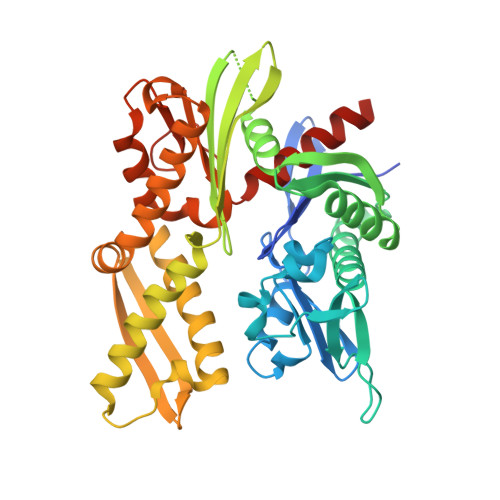
    4GMQ, 4GNI

  • PubMed Abstract: 

    Ribosome-associated chaperones act in early folding events during protein synthesis. Structural information is available for prokaryotic chaperones (such as trigger factor), but structural understanding of these processes in eukaryotes lags far behind. Here we present structural analyses of the eukaryotic ribosome-associated complex (RAC) from Saccharomyces cerevisiae and Chaetomium thermophilum, consisting of heat-shock protein 70 (Hsp70) Ssz1 and the Hsp40 Zuo1. RAC is an elongated complex that crouches over the ribosomal tunnel exit and seems to be stabilized in a distinct conformation by expansion segment ES27. A unique α-helical domain in Zuo1 mediates ribosome interaction of RAC near the ribosomal proteins L22e and L31e and ribosomal RNA helix H59. The crystal structure of the Ssz1 ATPase domain bound to ATP-Mg²⁺ explains its catalytic inactivity and suggests that Ssz1 may act before the RAC-associated chaperone Ssb. Our study offers insights into the interplay between RAC, the ER membrane-integrated Hsp40-type protein ERj1 and the signal-recognition particle.


  • Organizational Affiliation
    • Gene Center and Center of Integrated Protein Science Munich-CiPS-M, Department of Chemistry and Biochemistry, University of Munich, Munich, Germany.

Macromolecule Content 

  • Total Structure Weight: 89.97 kDa 
  • Atom Count: 6,783 
  • Modeled Residue Count: 769 
  • Deposited Residue Count: 818 
  • Unique protein chains: 1

Macromolecules

Find similar proteins by:|  3D Structure
Entity ID: 1
MoleculeChains  Sequence LengthOrganismDetailsImage
Putative heat shock protein
A, B
409Thermochaetoides thermophila DSM 1495Mutation(s): 0 
Gene Names: CTHT_0008010
UniProt
Find proteins for G0RZX9 (Chaetomium thermophilum (strain DSM 1495 / CBS 144.50 / IMI 039719))
Explore G0RZX9 
Go to UniProtKB:  G0RZX9
Entity Groups
Sequence Clusters30% Identity50% Identity70% Identity90% Identity95% Identity100% Identity
UniProt GroupG0RZX9
Sequence Annotations
Expand
Reference Sequence

Experimental Data & Validation

Experimental Data

  • Method: X-RAY DIFFRACTION
  • Resolution: 1.80 Å
  • R-Value Free:  0.244 (Depositor), 0.239 (DCC) 
  • R-Value Work:  0.201 (Depositor), 0.198 (DCC) 
  • R-Value Observed: 0.202 (Depositor) 
Space Group: P 1 21 1
Unit Cell:
Length ( Å )Angle ( ˚ )
a = 80.63α = 90
b = 55.38β = 106.53
c = 98.79γ = 90
Software Package:
Software NamePurpose
ADSCdata collection
PHASERphasing
PHENIXrefinement
MOSFLMdata reduction
SCALAdata scaling

Structure Validation

View Full Validation Report


Ligand Structure Quality Assessment 


Entry History 

Deposition Data

Revision History  (Full details and data files)

  • Version 1.0: 2012-12-05
    Type: Initial release
  • Version 1.1: 2013-01-02
    Changes: Database references
  • Version 1.2: 2013-01-16
    Changes: Database references
  • Version 1.3: 2024-02-28
    Changes: Data collection, Database references, Derived calculations