4GMG | pdb_00004gmg

NADP+ bound structure of a Thiazolinyl Imine Reductase from Yersinia enterocolitica (Irp3)


Experimental Data Snapshot

  • Method: X-RAY DIFFRACTION
  • Resolution: 2.31 Å
  • R-Value Free: 
    0.260 (Depositor), 0.258 (DCC) 
  • R-Value Work: 
    0.197 (Depositor), 0.196 (DCC) 
  • R-Value Observed: 
    0.200 (Depositor) 

wwPDB Validation 3D Report Full Report

Validation slider image for 4GMG

Ligand Structure Quality Assessment 


This is version 1.2 of the entry. See complete history

Literature

Two Structures of a Thiazolinyl Imine Reductase from Yersinia enterocolitica Provide Insight into Catalysis and Binding to the Nonribosomal Peptide Synthetase Module of HMWP1.

Meneely, K.M.Lamb, A.L.

(2012) Biochemistry 51: 9002-9013

  • DOI: https://doi.org/10.1021/bi3011016
  • Primary Citation Related Structures: 
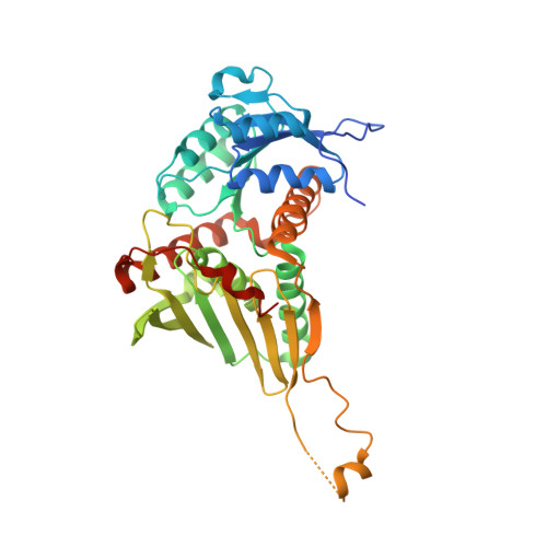
    4GMF, 4GMG

  • PubMed Abstract: 

    The thiazolinyl imine reductase from Yersinia enterocolitica (Irp3) catalyzes the NADPH-dependent reduction of a thiazoline ring in an intermediate for the formation of the siderophore yersiniabactin. Two structures of Irp3 were determined in the apo (1.85 Å) and NADP(+)-bound (2.31 Å) forms. Irp3 is structurally homologous to sugar oxidoreductases such as glucose-fructose oxidoreductase and 1,5-anhydro-d-fructose reductase, as well as to biliverdin reductase. A homology model of the thiazolinyl imine reductase from Pseudomonas aeruginosa (PchG) was generated. Extensive loop insertions are observed in the C-terminal domain that are unique to Irp3 and PchG and not found in the structural homologues that recognize small molecular substrates. These loops are hypothesized to be important for binding of the nonribosomal peptide synthetase modules (found in HMWP1 and PchF, respectively) to which the substrate of the reductase is covalently attached. A catalytic mechanism for the donation of a proton from a general acid (either histidine 101 or tyrosine 128) and the donation of a hydride from C4 of nicotinamide of the NADPH cofactor is proposed for reduction of the carbon-nitrogen double bond of the thiazoline.


  • Organizational Affiliation
    • Department of Molecular Biosciences, University of Kansas, Lawrence, Kansas 66045, United States.

Macromolecule Content 

  • Total Structure Weight: 174.68 kDa 
  • Atom Count: 11,156 
  • Modeled Residue Count: 1,382 
  • Deposited Residue Count: 1,540 
  • Unique protein chains: 1

Macromolecules

Find similar proteins by:|  3D Structure
Entity ID: 1
MoleculeChains  Sequence LengthOrganismDetailsImage
Yersiniabactin biosynthetic protein YbtU
A, B, C, D
385Yersinia enterocolitica subsp. enterocolitica 8081Mutation(s): 0 
Gene Names: Irp3ybtUYE2619
UniProt
Find proteins for A1JTG0 (Yersinia enterocolitica serotype O:8 / biotype 1B (strain NCTC 13174 / 8081))
Explore A1JTG0 
Go to UniProtKB:  A1JTG0
Entity Groups
Sequence Clusters30% Identity50% Identity70% Identity90% Identity95% Identity100% Identity
UniProt GroupA1JTG0
Sequence Annotations
Expand
Reference Sequence

Experimental Data & Validation

Experimental Data

  • Method: X-RAY DIFFRACTION
  • Resolution: 2.31 Å
  • R-Value Free:  0.260 (Depositor), 0.258 (DCC) 
  • R-Value Work:  0.197 (Depositor), 0.196 (DCC) 
  • R-Value Observed: 0.200 (Depositor) 
Space Group: P 21 21 21
Unit Cell:
Length ( Å )Angle ( ˚ )
a = 83.415α = 90
b = 93.817β = 90
c = 181.987γ = 90
Software Package:
Software NamePurpose
Blu-Icedata collection
PHASERphasing
REFMACrefinement
XDSdata reduction
SCALAdata scaling

Structure Validation

View Full Validation Report


Ligand Structure Quality Assessment 


Entry History 

Deposition Data

Revision History  (Full details and data files)

  • Version 1.0: 2012-10-31
    Type: Initial release
  • Version 1.1: 2012-12-12
    Changes: Database references
  • Version 1.2: 2024-02-28
    Changes: Data collection, Database references, Derived calculations