4GJ7

Crystal structure of renin in complex with NVP-BCA079 (compound 12a)


Experimental Data Snapshot

  • Method: X-RAY DIFFRACTION
  • Resolution: 2.80 Å
  • R-Value Free: 0.220 
  • R-Value Work: 0.180 
  • R-Value Observed: 0.184 

Starting Model: experimental
View more details

wwPDB Validation   3D Report Full Report


Ligand Structure Quality Assessment 


This is version 1.4 of the entry. See complete history


Literature

The Discovery of Novel Potent trans-3,4-Disubstituted Pyrrolidine Inhibitors of the Human Aspartic Protease Renin from in Silico Three-Dimensional (3D) Pharmacophore Searches.

Lorthiois, E.Breitenstein, W.Cumin, F.Ehrhardt, C.Francotte, E.Jacoby, E.Ostermann, N.Sellner, H.Kosaka, T.Webb, R.L.Rigel, D.F.Hassiepen, U.Richert, P.Wagner, T.Maibaum, J.

(2013) J Med Chem 56: 2207-2217

  • DOI: https://doi.org/10.1021/jm3017078
  • Primary Citation of Related Structures:  
    4GJ5, 4GJ6, 4GJ7

  • PubMed Abstract: 

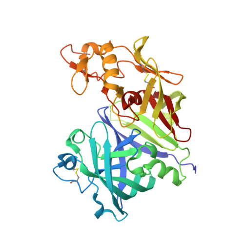
    The small-molecule trans-3,4-disubstituted pyrrolidine 6 was identified from in silico three-dimensional (3D) pharmacophore searches based on known X-ray structures of renin-inhibitor complexes and demonstrated to be a weakly active inhibitor of the human enzyme. The unexpected binding mode of the more potent enantiomer (3S,4S)-6a in an extended conformation spanning the nonprime and S1' pockets of the recombinant human (rh)-renin active site was elucidated by X-ray crystallography. Initial structure-activity relationship work focused on modifications of the hydrophobic diphenylamine portion positioned in S1 and extending toward the S2 pocket. Replacement with an optimized P3-P1 pharmacophore interacting to the nonsubstrate S3(sp) cavity eventually resulted in significantly improved in vitro potency and selectivity. The prototype analogue (3S,4S)-12a of this new class of direct renin inhibitors exerted blood pressure lowering effects in a hypertensive double-transgenic rat model after oral administration.


  • Organizational Affiliation

    Novartis Pharma AG, Institutes for BioMedical Research, Novartis Campus, CH-4056 Basel, Switzerland. edwige.lorthiois@novartis.com


Macromolecules
Find similar proteins by:  (by identity cutoff)  |  3D Structure
Entity ID: 1
MoleculeChains Sequence LengthOrganismDetailsImage
Renin
A, B
340Homo sapiensMutation(s): 0 
Gene Names: REN
EC: 3.4.23.15
UniProt & NIH Common Fund Data Resources
Find proteins for P00797 (Homo sapiens)
Explore P00797 
Go to UniProtKB:  P00797
GTEx:  ENSG00000143839 
Entity Groups  
Sequence Clusters30% Identity50% Identity70% Identity90% Identity95% Identity100% Identity
UniProt GroupP00797
Glycosylation
Glycosylation Sites: 1Go to GlyGen: P00797-1
Sequence Annotations
Expand
  • Reference Sequence
Small Molecules
Binding Affinity Annotations 
IDSourceBinding Affinity
0LT BindingDB:  4GJ7 IC50: min: 170, max: 450 (nM) from 3 assay(s)
PDBBind:  4GJ7 IC50: 170 (nM) from 1 assay(s)
Experimental Data & Validation

Experimental Data

  • Method: X-RAY DIFFRACTION
  • Resolution: 2.80 Å
  • R-Value Free: 0.220 
  • R-Value Work: 0.180 
  • R-Value Observed: 0.184 
  • Space Group: P 21 3
Unit Cell:
Length ( Å )Angle ( ˚ )
a = 141.476α = 90
b = 141.476β = 90
c = 141.476γ = 90
Software Package:
Software NamePurpose
BUSTERrefinement
DENZOdata reduction
SCALEPACKdata scaling

Structure Validation

View Full Validation Report


Ligand Structure Quality Assessment 


Entry History 

Deposition Data

Revision History  (Full details and data files)

  • Version 1.0: 2013-03-06
    Type: Initial release
  • Version 1.1: 2013-04-10
    Changes: Database references
  • Version 1.2: 2018-04-04
    Changes: Data collection
  • Version 1.3: 2020-07-29
    Type: Remediation
    Reason: Carbohydrate remediation
    Changes: Data collection, Derived calculations, Structure summary
  • Version 1.4: 2023-09-13
    Changes: Data collection, Database references, Refinement description, Structure summary