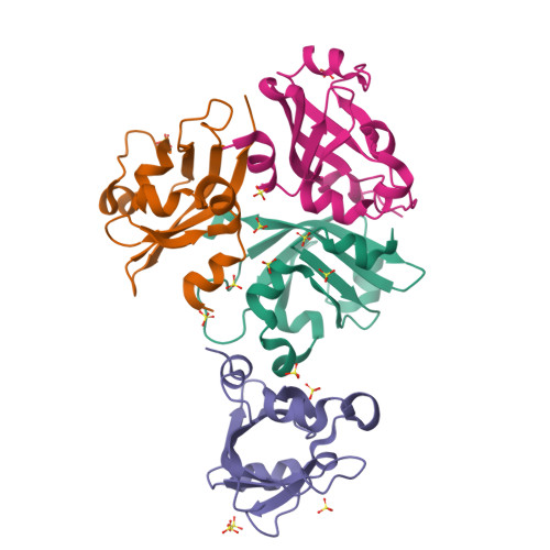
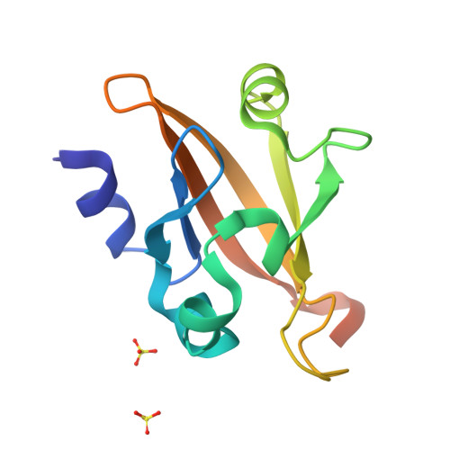
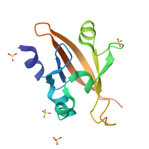
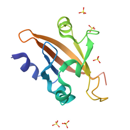
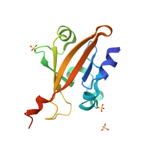
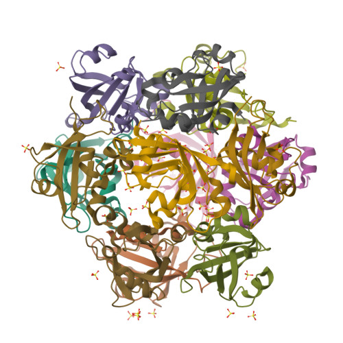
4GJ4

The Crystal Structure of the soluble Guanylate Cyclase PAS alpha domain from Manduca sexta


Experimental Data Snapshot

  • Method: X-RAY DIFFRACTION
  • Resolution: 1.80 Å
  • R-Value Free: 0.242 
  • R-Value Work: 0.196 
  • R-Value Observed: 0.198 

Starting Model: experimental
View more details

wwPDB Validation   3D Report Full Report


This is version 1.4 of the entry. See complete history


Literature

Crystal structure of the Alpha subunit PAS domain from soluble guanylyl cyclase.

Purohit, R.Weichsel, A.Montfort, W.R.

(2013) Protein Sci 22: 1439-1444

  • DOI: https://doi.org/10.1002/pro.2331
  • Primary Citation of Related Structures:  
    4GJ4

  • PubMed Abstract: 

    Soluble guanylate cyclase (sGC) is a heterodimeric heme protein of ≈ 150 kDa and the primary nitric oxide receptor. Binding of NO stimulates cyclase activity, leading to regulation of cardiovascular physiology and providing attractive opportunities for drug discovery. How sGC is stimulated and where candidate drugs bind remains unknown. The α and β sGC chains are each composed of Heme-Nitric Oxide Oxygen (H-NOX), Per-ARNT-Sim (PAS), coiled-coil and cyclase domains. Here, we present the crystal structure of the α1 PAS domain to 1.8 Å resolution. The structure reveals the binding surfaces of importance to heterodimer function, particularly with respect to regulating NO binding to heme in the β1 H-NOX domain. It also reveals a small internal cavity that may serve to bind ligands or participate in signal transduction.


  • Organizational Affiliation

    Department of Chemistry and Biochemistry, University of Arizona, Tucson, Arizona, 85721.


Macromolecules
Find similar proteins by:  (by identity cutoff)  |  3D Structure
Entity ID: 1
MoleculeChains Sequence LengthOrganismDetailsImage
Soluble guanylyl cyclase alpha-1 subunit
A, B, C, D
126Manduca sextaMutation(s): 3 
EC: 4.6.1.2
UniProt
Find proteins for O77105 (Manduca sexta)
Explore O77105 
Go to UniProtKB:  O77105
Entity Groups  
Sequence Clusters30% Identity50% Identity70% Identity90% Identity95% Identity100% Identity
UniProt GroupO77105
Sequence Annotations
Expand
  • Reference Sequence
Small Molecules
Ligands 1 Unique
IDChains Name / Formula / InChI Key2D Diagram3D Interactions
SO4
Query on SO4

Download Ideal Coordinates CCD File 
E [auth A]
F [auth B]
G [auth B]
H [auth B]
I [auth C]
E [auth A],
F [auth B],
G [auth B],
H [auth B],
I [auth C],
J [auth C],
K [auth C],
L [auth C],
M [auth D],
N [auth D],
O [auth D]
SULFATE ION
O4 S
QAOWNCQODCNURD-UHFFFAOYSA-L
Experimental Data & Validation

Experimental Data

  • Method: X-RAY DIFFRACTION
  • Resolution: 1.80 Å
  • R-Value Free: 0.242 
  • R-Value Work: 0.196 
  • R-Value Observed: 0.198 
  • Space Group: H 3 2
Unit Cell:
Length ( Å )Angle ( ˚ )
a = 95.424α = 90
b = 95.424β = 90
c = 317.693γ = 120
Software Package:
Software NamePurpose
MAR345data collection
MrBUMPphasing
REFMACrefinement
CrystalCleardata reduction
CrystalCleardata scaling

Structure Validation

View Full Validation Report


Entry History 

Deposition Data

Revision History  (Full details and data files)

  • Version 1.0: 2013-08-14
    Type: Initial release
  • Version 1.1: 2013-08-28
    Changes: Database references
  • Version 1.2: 2013-10-09
    Changes: Database references
  • Version 1.3: 2017-11-15
    Changes: Refinement description
  • Version 1.4: 2023-09-13
    Changes: Data collection, Database references, Derived calculations, Refinement description