4GIX

Crystal structure of human GLTP bound with 12:0 disulfatide


Experimental Data Snapshot

  • Method: X-RAY DIFFRACTION
  • Resolution: 1.80 Å
  • R-Value Free: 
    0.251 (Depositor), 0.190 (DCC) 
  • R-Value Work: 
    0.183 (Depositor), 0.190 (DCC) 
  • R-Value Observed: 
    0.187 (Depositor) 

Starting Model: experimental
View more details

wwPDB Validation   3D Report Full Report


Ligand Structure Quality Assessment 

Created with Raphaël 2.3.0Worse 01 BetterLigand structure goodness of fit to experimental dataBest fitted 0SGClick on this verticalbar to view details

This is version 1.1 of the entry. See complete history


Literature

Structural insights into lipid-dependent reversible dimerization of human GLTP.

Samygina, V.R.Ochoa-Lizarralde, B.Popov, A.N.Cabo-Bilbao, A.Goni-de-Cerio, F.Molotkovsky, J.G.Patel, D.J.Brown, R.E.Malinina, L.

(2013) Acta Crystallogr D Biol Crystallogr 69: 603-616

  • DOI: https://doi.org/10.1107/S0907444913000024
  • Primary Citation of Related Structures:  
    4GH0, 4GHP, 4GHS, 4GIX, 4GJQ, 4GVT, 4GXD, 4GXG, 4H2Z

  • PubMed Abstract: 

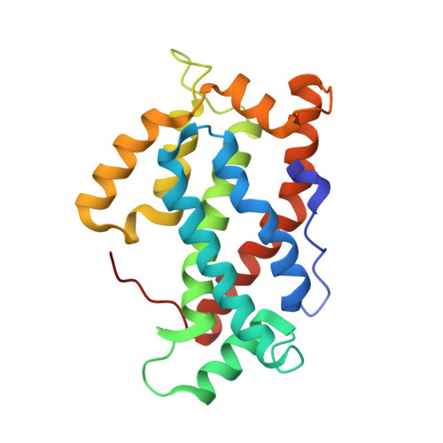
    Human glycolipid transfer protein (hsGLTP) forms the prototypical GLTP fold and is characterized by a broad transfer selectivity for glycosphingolipids (GSLs). The GLTP mutation D48V near the `portal entrance' of the glycolipid binding site has recently been shown to enhance selectivity for sulfatides (SFs) containing a long acyl chain. Here, nine novel crystal structures of hsGLTP and the SF-selective mutant complexed with short-acyl-chain monoSF and diSF in different crystal forms are reported in order to elucidate the potential functional roles of lipid-mediated homodimerization. In all crystal forms, the hsGLTP-SF complexes displayed homodimeric structures supported by similarly organized intermolecular interactions. The dimerization interface always involved the lipid sphingosine chain, the protein C-terminus (C-end) and α-helices 6 and 2, but the D48V mutant displayed a `locked' dimer conformation compared with the hinge-like flexibility of wild-type dimers. Differences in contact angles, areas and residues at the dimer interfaces in the `flexible' and `locked' dimers revealed a potentially important role of the dimeric structure in the C-end conformation of hsGLTP and in the precise positioning of the key residue of the glycolipid recognition centre, His140. ΔY207 and ΔC-end deletion mutants, in which the C-end is shifted or truncated, showed an almost complete loss of transfer activity. The new structural insights suggest that ligand-dependent reversible dimerization plays a role in the function of human GLTP.


  • Organizational Affiliation

    Structural Biology Unit, CIC bioGUNE, Technology Park of Bizkaia, 48160 Derio, Spain.


Macromolecules
Find similar proteins by:  (by identity cutoff)  |  3D Structure
Entity ID: 1
MoleculeChains Sequence LengthOrganismDetailsImage
Glycolipid transfer protein209Homo sapiensMutation(s): 0 
Gene Names: GLTP
UniProt & NIH Common Fund Data Resources
Find proteins for Q9NZD2 (Homo sapiens)
Explore Q9NZD2 
Go to UniProtKB:  Q9NZD2
PHAROS:  Q9NZD2
GTEx:  ENSG00000139433 
Entity Groups  
Sequence Clusters30% Identity50% Identity70% Identity90% Identity95% Identity100% Identity
UniProt GroupQ9NZD2
Sequence Annotations
Expand
  • Reference Sequence
Small Molecules
Ligands 1 Unique
IDChains Name / Formula / InChI Key2D Diagram3D Interactions
0SG
Query on 0SG

Download Ideal Coordinates CCD File 
B [auth A]N-{(2S,3R,4E)-1-[(3,6-di-O-sulfo-beta-D-galactopyranosyl)oxy]-3-hydroxyoctadec-4-en-2-yl}dodecanamide
C36 H69 N O14 S2
JBJSVLZKWGLXMO-AZNTWQPMSA-N
Experimental Data & Validation

Experimental Data

  • Method: X-RAY DIFFRACTION
  • Resolution: 1.80 Å
  • R-Value Free:  0.251 (Depositor), 0.190 (DCC) 
  • R-Value Work:  0.183 (Depositor), 0.190 (DCC) 
  • R-Value Observed: 0.187 (Depositor) 
Space Group: C 1 2 1
Unit Cell:
Length ( Å )Angle ( ˚ )
a = 74.768α = 90
b = 50.163β = 118.82
c = 65.85γ = 90
Software Package:
Software NamePurpose
MAR345dtbdata collection
MOLREPphasing
REFMACrefinement
HKL-2000data reduction
HKL-2000data scaling

Structure Validation

View Full Validation Report


Ligand Structure Quality Assessment 

Created with Raphaël 2.3.0Worse 01 BetterLigand structure goodness of fit to experimental dataBest fitted 0SGClick on this verticalbar to view details

Entry History 

Deposition Data

Revision History  (Full details and data files)

  • Version 1.0: 2013-04-10
    Type: Initial release
  • Version 1.1: 2023-11-08
    Changes: Data collection, Database references, Derived calculations, Refinement description