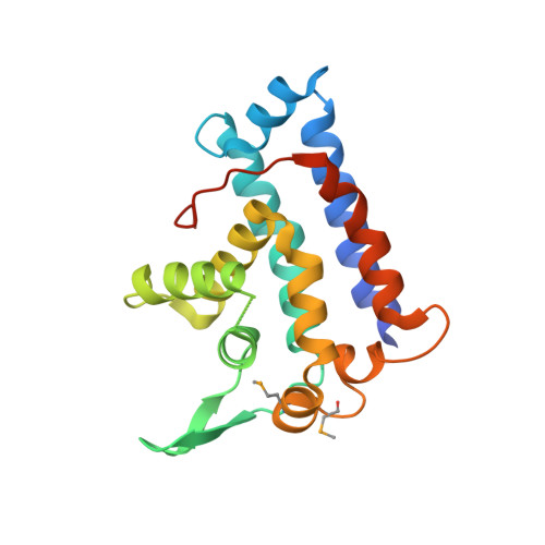
4GIW

Crystal structure of the RUN domain of human NESCA


Experimental Data Snapshot

  • Method: X-RAY DIFFRACTION
  • Resolution: 2.00 Å
  • R-Value Free: 0.212 
  • R-Value Work: 0.204 
  • R-Value Observed: 0.205 

wwPDB Validation   3D Report Full Report


This is version 1.1 of the entry. See complete history


Literature

Crystal structure and functional implication of the RUN domain of human NESCA

Sun, Q.Han, C.Liu, L.Wang, Y.Deng, H.Bai, L.Jiang, T.

(2012) Protein Cell 3: 609-617

  • DOI: https://doi.org/10.1007/s13238-012-2052-3
  • Primary Citation of Related Structures:  
    4GIW

  • PubMed Abstract: 

    NESCA, a newly discovered signaling adapter protein in the NGF-pathway, contains a RUN domain at its N-terminus. Here we report the crystal structure of the NESCA RUN domain determined at 2.0-Å resolution. The overall fold of the NESCA RUN domain comprises nine helices, resembling the RUN domain of RPIPx and the RUN1 domain of Rab6IP1. However, compared to the other RUN domains, the RUN domain of NESCA has significantly different surface electrostatic distributions at the putative GTPase-interacting interface. We demonstrate that the RUN domain of NESCA can bind H-Ras, a downstream signaling molecule of TrkA, with high affinity. Moreover, NESCA RUN can directly interact with TrkA. These results provide new insights into how NESCA participates in the NGF-TrkA signaling pathway.


  • Organizational Affiliation

    National Laboratory of Biomacromolecules, Institute of Biophysics, Chinese Academy of Sciences, Beijing, China.


Macromolecules
Find similar proteins by:  (by identity cutoff)  |  3D Structure
Entity ID: 1
MoleculeChains Sequence LengthOrganismDetailsImage
RUN and SH3 domain-containing protein 1
A, B
198Homo sapiensMutation(s): 2 
Gene Names: NESCA
UniProt & NIH Common Fund Data Resources
Find proteins for Q9BVN2 (Homo sapiens)
Explore Q9BVN2 
Go to UniProtKB:  Q9BVN2
PHAROS:  Q9BVN2
GTEx:  ENSG00000160753 
Entity Groups  
Sequence Clusters30% Identity50% Identity70% Identity90% Identity95% Identity100% Identity
UniProt GroupQ9BVN2
Sequence Annotations
Expand
  • Reference Sequence
Small Molecules
Modified Residues  1 Unique
IDChains TypeFormula2D DiagramParent
MSE
Query on MSE
A, B
L-PEPTIDE LINKINGC5 H11 N O2 SeMET
Experimental Data & Validation

Experimental Data

  • Method: X-RAY DIFFRACTION
  • Resolution: 2.00 Å
  • R-Value Free: 0.212 
  • R-Value Work: 0.204 
  • R-Value Observed: 0.205 
  • Space Group: P 1 21 1
Unit Cell:
Length ( Å )Angle ( ˚ )
a = 39.116α = 90
b = 94.201β = 99.54
c = 44.602γ = 90
Software Package:
Software NamePurpose
HKL-2000data collection
PHENIXmodel building
REFMACrefinement
HKL-2000data reduction
HKL-2000data scaling
PHENIXphasing

Structure Validation

View Full Validation Report


Entry History 

Deposition Data

Revision History  (Full details and data files)

  • Version 1.0: 2012-08-29
    Type: Initial release
  • Version 1.1: 2024-11-06
    Changes: Data collection, Database references, Derived calculations, Structure summary