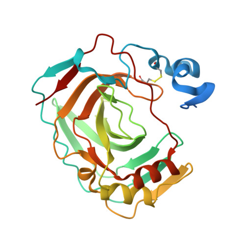
X-ray structure of the first `extremo-{alpha}-carbonic anhydrase', a dimeric enzyme from the thermophilic bacterium Sulfurihydrogenibium yellowstonense YO3AOP1.
Di Fiore, A., Capasso, C., De Luca, V., Monti, S.M., Carginale, V., Supuran, C.T., Scozzafava, A., Pedone, C., Rossi, M., De Simone, G.(2013) Acta Crystallogr D Biol Crystallogr 69: 1150-1159
- PubMed: 23695259 Search on PubMed
- DOI: https://doi.org/10.1107/S0907444913007208
- Primary Citation Related Structures:
4G7A - PubMed Abstract:
SspCA, a novel `extremo-α-carbonic anhydrase' isolated from the thermophilic bacterium Sulfurihydrogenibium yellowstonense YO3AOP1, is an efficient catalyst for the hydration of CO2 and presents exceptional thermostability. Indeed, SspCA retains a high catalytic activity even after being heated to 343-373 K for several hours. Here, the crystallographic structure of this α-carbonic anhydrase (α-CA) is reported and the factors responsible for its function at high temperature are elucidated. In particular, the study suggests that increased structural compactness, together with an increased number of charged residues on the protein surface and a greater number of ionic networks, seem to be the key factors involved in the higher thermostability of this enzyme with respect to its mesophilic homologues. These findings are of extreme importance, since they provide a structural basis for the understanding of the mechanisms responsible for thermal stability in the α-CA family for the first time. The data obtained offer a tool that can be exploited to engineer α-CAs in order to obtain enzymes with enhanced thermostability for use in the harsh conditions of the CO2 capture and sequestration processes.
- Istituto di Biostrutture e Bioimmagini - CNR, Via Mezzocannone 16, 80134 Napoli, Italy.
Organizational Affiliation: