4G6V | pdb_00004g6v

CdiA-CT/CdiI toxin and immunity complex from Burkholderia pseudomallei


Experimental Data Snapshot

  • Method: X-RAY DIFFRACTION
  • Resolution: 2.64 Å
  • R-Value Free: 
    0.245 (Depositor), 0.233 (DCC) 
  • R-Value Work: 
    0.204 (Depositor), 0.194 (DCC) 
  • R-Value Observed: 
    0.206 (Depositor) 

wwPDB Validation 3D Report Full Report

Validation slider image for 4G6V

This is version 1.2 of the entry. See complete history

Literature

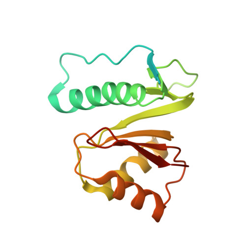
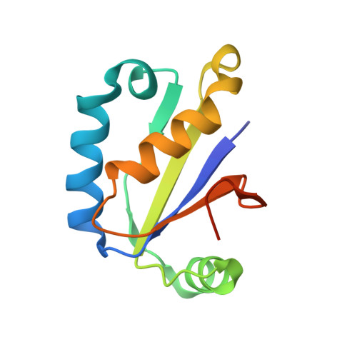
Structural basis of toxicity and immunity in contact-dependent growth inhibition (CDI) systems.

Morse, R.P.Nikolakakis, K.C.Willett, J.L.Gerrick, E.Low, D.A.Hayes, C.S.Goulding, C.W.

(2012) Proc Natl Acad Sci U S A 109: 21480-21485

  • DOI: https://doi.org/10.1073/pnas.1216238110
  • Primary Citation Related Structures: 
    4G6U, 4G6V

  • PubMed Abstract: 

    Contact-dependent growth inhibition (CDI) systems encode polymorphic toxin/immunity proteins that mediate competition between neighboring bacterial cells. We present crystal structures of CDI toxin/immunity complexes from Escherichia coli EC869 and Burkholderia pseudomallei 1026b. Despite sharing little sequence identity, the toxin domains are structurally similar and have homology to endonucleases. The EC869 toxin is a Zn(2+)-dependent DNase capable of completely degrading the genomes of target cells, whereas the Bp1026b toxin cleaves the aminoacyl acceptor stems of tRNA molecules. Each immunity protein binds and inactivates its cognate toxin in a unique manner. The EC869 toxin/immunity complex is stabilized through an unusual β-augmentation interaction. In contrast, the Bp1026b immunity protein exploits shape and charge complementarity to occlude the toxin active site. These structures represent the initial glimpse into the CDI toxin/immunity network, illustrating how sequence-diverse toxins adopt convergent folds yet retain distinct binding interactions with cognate immunity proteins. Moreover, we present visual demonstration of CDI toxin delivery into a target cell.


  • Organizational Affiliation
    • Department of Molecular Biology and Biochemistry, University of California, Irvine, CA 92697, USA.

Macromolecule Content 

  • Total Structure Weight: 123.45 kDa 
  • Atom Count: 6,969 
  • Modeled Residue Count: 936 
  • Deposited Residue Count: 1,148 
  • Unique protein chains: 2

Macromolecules

Find similar proteins by:|  3D Structure
Entity ID: 1
MoleculeChains  Sequence LengthOrganismDetailsImage
Adhesin/hemolysin
A, C, E, G
176Burkholderia pseudomallei 1026aMutation(s): 0 
Gene Names: BP1026A_3896
Find similar proteins by:|  3D Structure
Entity ID: 2
MoleculeChains  Sequence LengthOrganismDetailsImage
CdiI
B, D, F, H
111Burkholderia pseudomallei 1026aMutation(s): 0 
Gene Names: cdiI
UniProt
Find proteins for H9T8H3 (Burkholderia pseudomallei)
Explore H9T8H3 
Go to UniProtKB:  H9T8H3
Entity Groups
Sequence Clusters30% Identity50% Identity70% Identity90% Identity95% Identity100% Identity
UniProt GroupH9T8H3
Sequence Annotations
Expand
Reference Sequence

Experimental Data & Validation

Experimental Data

  • Method: X-RAY DIFFRACTION
  • Resolution: 2.64 Å
  • R-Value Free:  0.245 (Depositor), 0.233 (DCC) 
  • R-Value Work:  0.204 (Depositor), 0.194 (DCC) 
  • R-Value Observed: 0.206 (Depositor) 
Space Group: F 2 2 2
Unit Cell:
Length ( Å )Angle ( ˚ )
a = 151.963α = 90
b = 173.654β = 90
c = 174.822γ = 90
Software Package:
Software NamePurpose
HKL-2000data collection
PHENIXmodel building
PHENIXrefinement
HKL-2000data reduction
HKL-2000data scaling
PHENIXphasing

Structure Validation

View Full Validation Report


Entry History 

Deposition Data

Revision History  (Full details and data files)

  • Version 1.0: 2012-12-12
    Type: Initial release
  • Version 1.1: 2013-01-09
    Changes: Database references
  • Version 1.2: 2024-02-28
    Changes: Data collection, Database references, Derived calculations