4G2V | pdb_00004g2v

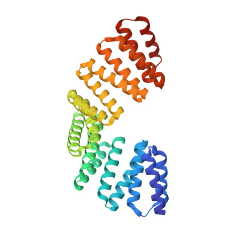
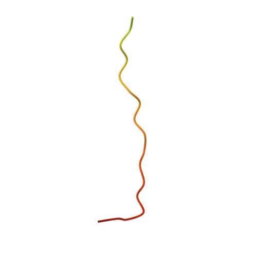
Structure complex of LGN binding with FRMPD1


Experimental Data Snapshot

  • Method: X-RAY DIFFRACTION
  • Resolution: 2.40 Å
  • R-Value Free: 
    0.232 (Depositor), 0.234 (DCC) 
  • R-Value Work: 
    0.190 (Depositor), 0.193 (DCC) 
  • R-Value Observed: 
    0.192 (Depositor) 

wwPDB Validation 3D Report Full Report

Validation slider image for 4G2V

This is version 1.2 of the entry. See complete history

Literature

Structural and biochemical characterization of the interaction between LGN and Frmpd1

Pan, Z.Shang, Y.Jia, M.Zhang, L.Xia, C.Zhang, M.Wang, W.Wen, W.

(2013) J Mol Biology 425: 1039-1049

  • DOI: https://doi.org/10.1016/j.jmb.2013.01.003
  • Primary Citation Related Structures: 
    4G2V

  • PubMed Abstract: 

    The tetratricopeptide repeat (TPR) motif-containing protein LGN binds multiple targets and regulates their subcellular localizations and functions during both asymmetric and symmetric cell divisions. Here, we characterized the interaction between LGN-TPR motifs and FERM and PDZ domain containing 1 (Frmpd1) and reported the crystal structure of the complex at 2.4Å resolution. A highly conserved fragment at the center of Frmpd1 of ~20 residues was found to be necessary and sufficient to bind to LGN-TPR. This Frmpd1 fragment forms an extended structure and runs along the concave channel of the TPR superhelix in an antiparallel manner in the complex. Structural comparisons and biochemical studies of LGN/Frmpd1 and other known LGN/target interactions demonstrate that the LGN-TPR motifs are versatile and capable of recognizing multiple targets via diverse binding modes. Nevertheless, a conserved "E/QxEx4-5E/D/Qx1-2K/R" motif in LGN/Pins (partner of inscuteable) TPR binding proteins has been identified.


  • Organizational Affiliation
    • Key Laboratory of Molecular Medicine, Ministry of Education, Department of Biochemistry and Molecular Biology, and Institutes of Biomedical Sciences, Shanghai Medical College, Fudan University, Shanghai 200032, P. R. China.

Macromolecule Content 

  • Total Structure Weight: 42.67 kDa 
  • Atom Count: 2,732 
  • Modeled Residue Count: 338 
  • Deposited Residue Count: 378 
  • Unique protein chains: 2

Macromolecules

Find similar proteins by:|  3D Structure
Entity ID: 1
MoleculeChains  Sequence LengthOrganismDetailsImage
G-protein-signaling modulator 2340Mus musculusMutation(s): 0 
UniProt & NIH Common Fund Data Resources
Find proteins for Q8VDU0 (Mus musculus)
Explore Q8VDU0 
Go to UniProtKB:  Q8VDU0
IMPC:  MGI:1923373
Entity Groups
Sequence Clusters30% Identity50% Identity70% Identity90% Identity95% Identity100% Identity
UniProt GroupQ8VDU0
Sequence Annotations
Expand
Reference Sequence
Find similar proteins by:|  3D Structure
Entity ID: 2
MoleculeChains  Sequence LengthOrganismDetailsImage
peptide from FERM and PDZ domain-containing protein 138Homo sapiensMutation(s): 0 
UniProt & NIH Common Fund Data Resources
Find proteins for Q5SYB0 (Homo sapiens)
Explore Q5SYB0 
Go to UniProtKB:  Q5SYB0
PHAROS:  Q5SYB0
GTEx:  ENSG00000070601 
Entity Groups
Sequence Clusters30% Identity50% Identity70% Identity90% Identity95% Identity100% Identity
UniProt GroupQ5SYB0
Sequence Annotations
Expand
Reference Sequence

Small Molecules

Ligands 3 Unique
IDChains Name / Formula / InChI Key2D Diagram3D Interactions
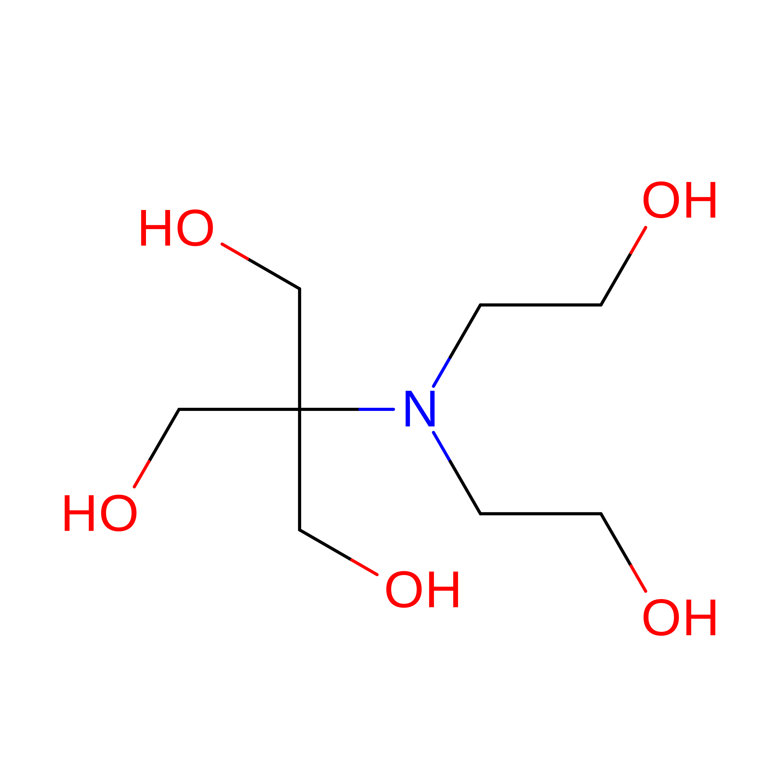
BTB

Query on BTB


Download:Ideal Coordinates CCD File
J [auth A]2-[BIS-(2-HYDROXY-ETHYL)-AMINO]-2-HYDROXYMETHYL-PROPANE-1,3-DIOL
C8 H19 N O5
OWMVSZAMULFTJU-UHFFFAOYSA-N
GOL

Query on GOL


Download:Ideal Coordinates CCD File
C [auth A]
D [auth A]
E [auth A]
F [auth A]
G [auth A]
C [auth A],
D [auth A],
E [auth A],
F [auth A],
G [auth A],
H [auth A],
I [auth A],
L [auth B]
GLYCEROL
C3 H8 O3
PEDCQBHIVMGVHV-UHFFFAOYSA-N
CL

Query on CL


Download:Ideal Coordinates CCD File
K [auth A]CHLORIDE ION
Cl
VEXZGXHMUGYJMC-UHFFFAOYSA-M

Experimental Data & Validation

Experimental Data

  • Method: X-RAY DIFFRACTION
  • Resolution: 2.40 Å
  • R-Value Free:  0.232 (Depositor), 0.234 (DCC) 
  • R-Value Work:  0.190 (Depositor), 0.193 (DCC) 
  • R-Value Observed: 0.192 (Depositor) 
Space Group: P 61 2 2
Unit Cell:
Length ( Å )Angle ( ˚ )
a = 93.835α = 90
b = 93.835β = 90
c = 172.863γ = 120
Software Package:
Software NamePurpose
HKL-2000data collection
PHASESphasing
PHENIXrefinement
HKL-2000data reduction
HKL-2000data scaling

Structure Validation

View Full Validation Report


Entry History 

Deposition Data

Revision History  (Full details and data files)

  • Version 1.0: 2013-01-23
    Type: Initial release
  • Version 1.1: 2013-07-17
    Changes: Database references
  • Version 1.2: 2024-03-20
    Changes: Data collection, Database references, Derived calculations