4G2C | pdb_00004g2c

DyP2 from Amycolatopsis sp. ATCC 39116


Experimental Data Snapshot

  • Method: X-RAY DIFFRACTION
  • Resolution: 2.25 Å
  • R-Value Free: 
    0.243 (Depositor), 0.209 (DCC) 
  • R-Value Work: 
    0.184 (Depositor) 
  • R-Value Observed: 
    0.187 (Depositor) 

Starting Model: experimental
View more details

wwPDB Validation 3D Report Full Report

Validation slider image for 4G2C

Ligand Structure Quality Assessment 


This is version 1.2 of the entry. See complete history

Literature

Identification and characterization of a multifunctional dye peroxidase from a lignin-reactive bacterium.

Brown, M.E.Barros, T.Chang, M.C.

(2012) ACS Chem Biol 7: 2074-2081

  • DOI: https://doi.org/10.1021/cb300383y
  • Primary Citation Related Structures: 
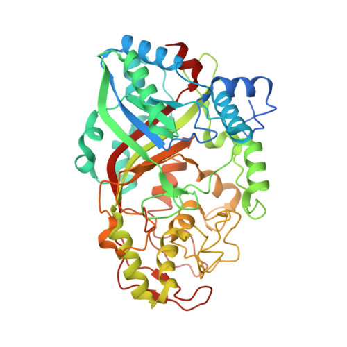
    4G2C

  • PubMed Abstract: 

    Plant biomass represents a renewable feedstock that has not yet been fully tapped because of the difficulty in accessing the carbon in its structural biopolymers. Lignin is an especially challenging substrate, but select microbes have evolved complex systems of enzymes for its breakdown through a radical-mediated oxidation process. Fungal systems are well-characterized for their ability to depolymerize lignin, but the ability of bacteria to react with this substrate remains elusive. We have therefore focused on elucidating strategies used by lignin-reactive soil bacteria and describing their oxidative enzyme systems. We now report the identification and characterization of an unusual C-type dye-decolorizing peroxidase from Amycolatopsis sp. 75iv2 (DyP2), which belongs to a family of heme peroxidases reported to be involved in bacterial lignin degradation. Biochemical studies indicate that DyP2 has novel function for this family, with versatile and high activity both as a peroxidase and Mn peroxidase (k(cat)/K(M) ≈ 10(5)-10(6) M(-1) s(-1)). It also has a Mn-dependent oxidase mode of action that expands its substrate scope. Crystallographic studies of DyP2 at 2.25 Å resolution show the existence of a Mn binding pocket and support its key role in catalysis.


  • Organizational Affiliation
    • Department of Chemistry, University of California, Berkeley, CA 94720-1460, USA.

Macromolecule Content 

  • Total Structure Weight: 110.63 kDa 
  • Atom Count: 7,712 
  • Modeled Residue Count: 925 
  • Deposited Residue Count: 986 
  • Unique protein chains: 1

Macromolecules

Find similar proteins by:|  3D Structure
Entity ID: 1
MoleculeChains  Sequence LengthOrganismDetailsImage
DyP2
A, B
493Amycolatopsis sp. ATCC 39116Mutation(s): 0 
EC: 1 (PDB Primary Data), 1.11.1.19 (UniProt), 1.11.1.7 (UniProt), 1.11.1.16 (UniProt)
UniProt
Find proteins for K7N5M8 (Amycolatopsis sp. (strain ATCC 39116 / 75iv2))
Explore K7N5M8 
Go to UniProtKB:  K7N5M8
Entity Groups
Sequence Clusters30% Identity50% Identity70% Identity90% Identity95% Identity100% Identity
UniProt GroupK7N5M8
Sequence Annotations
Expand
Reference Sequence

Small Molecules

Ligands 3 Unique
IDChains Name / Formula / InChI Key2D Diagram3D Interactions
HEM

Query on HEM


Download:Ideal Coordinates CCD File
C [auth A],
DA [auth B]
PROTOPORPHYRIN IX CONTAINING FE
C34 H32 Fe N4 O4
KABFMIBPWCXCRK-RGGAHWMASA-L
ACT

Query on ACT


Download:Ideal Coordinates CCD File
AA [auth A]
BA [auth A]
CA [auth A]
E [auth A]
F [auth A]
AA [auth A],
BA [auth A],
CA [auth A],
E [auth A],
F [auth A],
FA [auth B],
G [auth A],
GA [auth B],
H [auth A],
HA [auth B],
I [auth A],
IA [auth B],
J [auth A],
JA [auth B],
K [auth A],
KA [auth B],
L [auth A],
LA [auth B],
M [auth A],
MA [auth B],
N [auth A],
NA [auth B],
O [auth A],
OA [auth B],
P [auth A],
PA [auth B],
Q [auth A],
QA [auth B],
R [auth A],
RA [auth B],
S [auth A],
T [auth A],
U [auth A],
V [auth A],
W [auth A],
X [auth A],
Y [auth A],
Z [auth A]
ACETATE ION
C2 H3 O2
QTBSBXVTEAMEQO-UHFFFAOYSA-M
MN

Query on MN


Download:Ideal Coordinates CCD File
D [auth A],
EA [auth B]
MANGANESE (II) ION
Mn
WAEMQWOKJMHJLA-UHFFFAOYSA-N

Experimental Data & Validation

Experimental Data

  • Method: X-RAY DIFFRACTION
  • Resolution: 2.25 Å
  • R-Value Free:  0.243 (Depositor), 0.209 (DCC) 
  • R-Value Work:  0.184 (Depositor) 
  • R-Value Observed: 0.187 (Depositor) 
Space Group: P 21 21 21
Unit Cell:
Length ( Å )Angle ( ˚ )
a = 48.131α = 90
b = 77.952β = 90
c = 252.353γ = 90
Software Package:
Software NamePurpose
ADSCdata collection
PHASERphasing
PHENIXrefinement
XDSdata reduction
SCALAdata scaling

Structure Validation

View Full Validation Report


Ligand Structure Quality Assessment 


Entry History 

Deposition Data

Revision History  (Full details and data files)

  • Version 1.0: 2012-10-24
    Type: Initial release
  • Version 1.1: 2013-01-02
    Changes: Database references
  • Version 1.2: 2023-09-13
    Changes: Data collection, Database references, Derived calculations, Refinement description