4G0N | pdb_00004g0n

Crystal Structure of wt H-Ras-GppNHp bound to the RBD of Raf Kinase


Experimental Data Snapshot

  • Method: X-RAY DIFFRACTION
  • Resolution: 2.45 Å
  • R-Value Free: 
    0.229 (Depositor), 0.230 (DCC) 
  • R-Value Work: 
    0.181 (Depositor), 0.182 (DCC) 
  • R-Value Observed: 
    0.186 (Depositor) 

wwPDB Validation 3D Report Full Report

Validation slider image for 4G0N

Ligand Structure Quality Assessment 


This is version 1.2 of the entry. See complete history

Literature

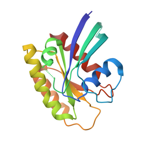
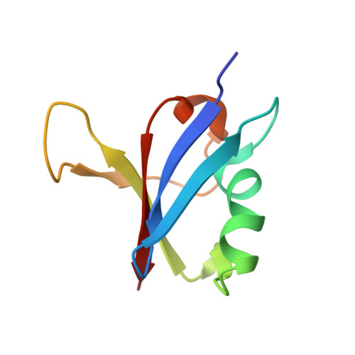
Allosteric Effects of the Oncogenic RasQ61L Mutant on Raf-RBD.

Fetics, S.K.Guterres, H.Kearney, B.M.Buhrman, G.Ma, B.Nussinov, R.Mattos, C.

(2015) Structure 23: 505-516

  • DOI: https://doi.org/10.1016/j.str.2014.12.017
  • Primary Citation Related Structures: 
    4G0N, 4G3X

  • PubMed Abstract: 

    The Ras/Raf/MEK/ERK signal transduction pathway is a major regulator of cell proliferation activated by Ras-guanosine triphosphate (GTP). The oncogenic mutant RasQ61L is not able to hydrolyze GTP in the presence of Raf and thus is a constitutive activator of this mitogenic pathway. The Ras/Raf interaction is essential for the activation of the Raf kinase domain through a currently unknown mechanism. We present the crystal structures of the Ras-GppNHp/Raf-RBD and RasQ61L-GppNHp/Raf-RBD complexes, which, in combination with MD simulations, reveal differences in allosteric interactions leading from the Ras/Raf interface to the Ras calcium-binding site and to the remote Raf-RBD loop L4. In the presence of Raf, the RasQ61L mutant has a rigid switch II relative to the wild-type and increased flexibility at the interface with switch I, which propagates across Raf-RBD. We show that in addition to local perturbations on Ras, RasQ61L has substantial long-range effects on the Ras allosteric lobe and on Raf-RBD.


  • Organizational Affiliation
    • Department of Chemistry and Chemical Biology, Northeastern University, Boston, MA 02115, USA; Department of Molecular and Structural Biochemistry, North Carolina State University, Raleigh, NC 27695, USA.

Macromolecule Content 

  • Total Structure Weight: 28.61 kDa 
  • Atom Count: 2,004 
  • Modeled Residue Count: 242 
  • Deposited Residue Count: 244 
  • Unique protein chains: 2

Macromolecules

Find similar proteins by:|  3D Structure
Entity ID: 1
MoleculeChains  Sequence LengthOrganismDetailsImage
GTPase HRas166Homo sapiensMutation(s): 0 
Gene Names: HRASHRAS1
EC: 3.6.5.2
UniProt & NIH Common Fund Data Resources
Find proteins for P01112 (Homo sapiens)
Explore P01112 
Go to UniProtKB:  P01112
PHAROS:  P01112
GTEx:  ENSG00000174775 
Entity Groups
Sequence Clusters30% Identity50% Identity70% Identity90% Identity95% Identity100% Identity
UniProt GroupP01112
Sequence Annotations
Expand
Reference Sequence
Find similar proteins by:|  3D Structure
Entity ID: 2
MoleculeChains  Sequence LengthOrganismDetailsImage
RAF proto-oncogene serine/threonine-protein kinase78Homo sapiensMutation(s): 0 
Gene Names: RAF1RAF
EC: 2.7.11.1
UniProt & NIH Common Fund Data Resources
Find proteins for P04049 (Homo sapiens)
Explore P04049 
Go to UniProtKB:  P04049
PHAROS:  P04049
GTEx:  ENSG00000132155 
Entity Groups
Sequence Clusters30% Identity50% Identity70% Identity90% Identity95% Identity100% Identity
UniProt GroupP04049
Sequence Annotations
Expand
Reference Sequence

Small Molecules

Ligands 5 Unique
IDChains Name / Formula / InChI Key2D Diagram3D Interactions
GNP

Query on GNP


Download:Ideal Coordinates CCD File
C [auth A]PHOSPHOAMINOPHOSPHONIC ACID-GUANYLATE ESTER
C10 H17 N6 O13 P3
UQABYHGXWYXDTK-UUOKFMHZSA-N
DTU

Query on DTU


Download:Ideal Coordinates CCD File
H [auth A](2R,3S)-1,4-DIMERCAPTOBUTANE-2,3-DIOL
C4 H10 O2 S2
VHJLVAABSRFDPM-ZXZARUISSA-N
ACT

Query on ACT


Download:Ideal Coordinates CCD File
G [auth A]ACETATE ION
C2 H3 O2
QTBSBXVTEAMEQO-UHFFFAOYSA-M
CA

Query on CA


Download:Ideal Coordinates CCD File
D [auth A],
F [auth A]
CALCIUM ION
Ca
BHPQYMZQTOCNFJ-UHFFFAOYSA-N
MG

Query on MG


Download:Ideal Coordinates CCD File
E [auth A]MAGNESIUM ION
Mg
JLVVSXFLKOJNIY-UHFFFAOYSA-N

Experimental Data & Validation

Experimental Data

  • Method: X-RAY DIFFRACTION
  • Resolution: 2.45 Å
  • R-Value Free:  0.229 (Depositor), 0.230 (DCC) 
  • R-Value Work:  0.181 (Depositor), 0.182 (DCC) 
  • R-Value Observed: 0.186 (Depositor) 
Space Group: P 3 2 1
Unit Cell:
Length ( Å )Angle ( ˚ )
a = 90.44α = 90
b = 90.44β = 90
c = 92.703γ = 120
Software Package:
Software NamePurpose
PHENIXmodel building
PHENIXrefinement
HKL-2000data reduction
HKL-2000data scaling
PHENIXphasing

Structure Validation

View Full Validation Report


Ligand Structure Quality Assessment 


Entry History 

Deposition Data

Revision History  (Full details and data files)

  • Version 1.0: 2013-07-17
    Type: Initial release
  • Version 1.1: 2015-03-11
    Changes: Database references
  • Version 1.2: 2024-02-28
    Changes: Data collection, Database references, Derived calculations