4FYR | pdb_00004fyr

Human aminopeptidase N (CD13) in complex with bestatin


Experimental Data Snapshot

  • Method: X-RAY DIFFRACTION
  • Resolution: 1.91 Å
  • R-Value Free: 
    0.190 (Depositor), 0.189 (DCC) 
  • R-Value Work: 
    0.167 (Depositor), 0.167 (DCC) 
  • R-Value Observed: 
    0.168 (Depositor) 

wwPDB Validation 3D Report Full Report

Validation slider image for 4FYR

Ligand Structure Quality Assessment 


This is version 2.1 of the entry. See complete history

Literature

The X-ray Crystal Structure of Human Aminopeptidase N Reveals a Novel Dimer and the Basis for Peptide Processing.

Wong, A.H.Zhou, D.Rini, J.M.

(2012) J Biological Chem 287: 36804-36813

  • DOI: https://doi.org/10.1074/jbc.M112.398842
  • Primary Citation Related Structures: 
    4FYQ, 4FYR, 4FYS, 4FYT

  • PubMed Abstract: 

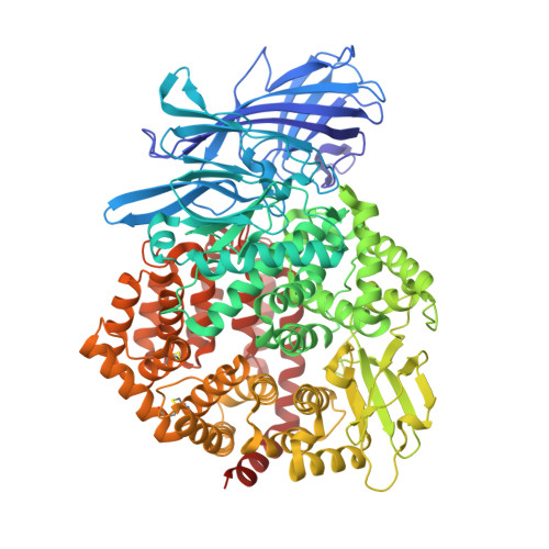
    Human aminopeptidase N (hAPN/hCD13) is a dimeric membrane protein and a member of the M1 family of zinc metallopeptidases. Within the rennin-angiotensin system, its enzymatic activity is responsible for processing peptide hormones angiotensin III and IV. In addition, hAPN is also involved in cell adhesion, endocytosis, and signal transduction and it is an important target for cancer therapy. Reported here are the high resolution x-ray crystal structures of the dimeric ectodomain of hAPN and its complexes with angiotensin IV and the peptidomimetic inhibitors, amastatin and bestatin. Each monomer of the dimer is found in what has been termed the closed form in other M1 enzymes and each monomer is characterized by an internal cavity surrounding the catalytic site as well as a unique substrate/inhibitor-dependent loop ordering, which in the case of the bestatin complex suggests a new route to inhibitor design. The hAPN structure provides the first example of a dimeric M1 family member and the observed structural features, in conjunction with a model for the open form, provide novel insights into the mechanism of peptide processing and signal transduction.


  • Organizational Affiliation
    • Department of Biochemistry, University of Toronto, Toronto, Ontario M5S 1A8, Canada.

Macromolecule Content 

  • Total Structure Weight: 107.2 kDa 
  • Atom Count: 8,543 
  • Modeled Residue Count: 903 
  • Deposited Residue Count: 903 
  • Unique protein chains: 1

Macromolecules

Find similar proteins by:|  3D Structure
Entity ID: 1
MoleculeChains  Sequence LengthOrganismDetailsImage
Aminopeptidase N903Homo sapiensMutation(s): 0 
Gene Names: ANPEPAPNCD13PEPN
EC: 3.4.11.2
UniProt & NIH Common Fund Data Resources
Find proteins for P15144 (Homo sapiens)
Explore P15144 
Go to UniProtKB:  P15144
PHAROS:  P15144
GTEx:  ENSG00000166825 
Entity Groups
Sequence Clusters30% Identity50% Identity70% Identity90% Identity95% Identity100% Identity
UniProt GroupP15144
Glycosylation
Glycosylation Sites: 9Go to GlyGen: P15144-1
Sequence Annotations
Expand
Reference Sequence

Oligosaccharides

Help  
Entity ID: 2
MoleculeChains Length2D Diagram GlycosylationD Interactions
beta-D-mannopyranose-(1-4)-2-acetamido-2-deoxy-beta-D-glucopyranose-(1-4)-2-acetamido-2-deoxy-beta-D-glucopyranose
B
3N-Glycosylation
Glycosylation Resources
GlyTouCan: G15407YE
GlyCosmos: G15407YE
GlyGen: G15407YE
Entity ID: 3
MoleculeChains Length2D Diagram GlycosylationD Interactions
2-acetamido-2-deoxy-beta-D-glucopyranose-(1-4)-2-acetamido-2-deoxy-beta-D-glucopyranose
C
2N-Glycosylation
Glycosylation Resources
GlyTouCan: G42666HT
GlyCosmos: G42666HT
GlyGen: G42666HT

Small Molecules

Ligands 5 Unique
IDChains Name / Formula / InChI Key2D Diagram3D Interactions
BES

Query on BES


Download:Ideal Coordinates CCD File
L [auth A]2-(3-AMINO-2-HYDROXY-4-PHENYL-BUTYRYLAMINO)-4-METHYL-PENTANOIC ACID
C16 H24 N2 O4
VGGGPCQERPFHOB-RDBSUJKOSA-N
NAG

Query on NAG


Download:Ideal Coordinates CCD File
D [auth A]
E [auth A]
F [auth A]
G [auth A]
H [auth A]
D [auth A],
E [auth A],
F [auth A],
G [auth A],
H [auth A],
I [auth A],
J [auth A]
2-acetamido-2-deoxy-beta-D-glucopyranose
C8 H15 N O6
OVRNDRQMDRJTHS-FMDGEEDCSA-N
SO4

Query on SO4


Download:Ideal Coordinates CCD File
M [auth A]
N [auth A]
O [auth A]
P [auth A]
Q [auth A]
M [auth A],
N [auth A],
O [auth A],
P [auth A],
Q [auth A],
R [auth A],
S [auth A],
T [auth A],
U [auth A],
V [auth A]
SULFATE ION
O4 S
QAOWNCQODCNURD-UHFFFAOYSA-L
ZN

Query on ZN


Download:Ideal Coordinates CCD File
K [auth A]ZINC ION
Zn
PTFCDOFLOPIGGS-UHFFFAOYSA-N
ACY

Query on ACY


Download:Ideal Coordinates CCD File
W [auth A]ACETIC ACID
C2 H4 O2
QTBSBXVTEAMEQO-UHFFFAOYSA-N

Experimental Data & Validation

Experimental Data

  • Method: X-RAY DIFFRACTION
  • Resolution: 1.91 Å
  • R-Value Free:  0.190 (Depositor), 0.189 (DCC) 
  • R-Value Work:  0.167 (Depositor), 0.167 (DCC) 
  • R-Value Observed: 0.168 (Depositor) 
Space Group: P 64
Unit Cell:
Length ( Å )Angle ( ˚ )
a = 158.068α = 90
b = 158.068β = 90
c = 114.809γ = 120
Software Package:
Software NamePurpose
DENZOdata reduction
SCALEPACKdata scaling
REFMACrefinement
PDB_EXTRACTdata extraction

Structure Validation

View Full Validation Report


Ligand Structure Quality Assessment 


Entry History 

Deposition Data

Revision History  (Full details and data files)

  • Version 1.0: 2012-09-05
    Type: Initial release
  • Version 1.1: 2012-11-14
    Changes: Database references
  • Version 1.2: 2017-11-15
    Changes: Refinement description
  • Version 2.0: 2020-07-29
    Type: Remediation
    Reason: Carbohydrate remediation
    Changes: Atomic model, Data collection, Database references, Derived calculations, Structure summary
  • Version 2.1: 2024-11-06
    Changes: Data collection, Database references, Structure summary