4FR8 | pdb_00004fr8

Crystal structure of human aldehyde dehydrogenase-2 in complex with nitroglycerin


Experimental Data Snapshot

  • Method: X-RAY DIFFRACTION
  • Resolution: 2.20 Å
  • R-Value Free: 
    0.167 (Depositor), 0.151 (DCC) 
  • R-Value Work: 
    0.132 (Depositor), 0.130 (DCC) 
  • R-Value Observed: 
    0.134 (Depositor) 

wwPDB Validation 3D Report Full Report

Validation slider image for 4FR8

Ligand Structure Quality Assessment 


This is version 1.2 of the entry. See complete history

Literature

Vascular Bioactivation of Nitroglycerin by Aldehyde Dehydrogenase-2: REACTION INTERMEDIATES REVEALED BY CRYSTALLOGRAPHY AND MASS SPECTROMETRY.

Lang, B.S.Gorren, A.C.Oberdorfer, G.Wenzl, M.V.Furdui, C.M.Poole, L.B.Mayer, B.Gruber, K.

(2012) J Biological Chem 287: 38124-38134

  • DOI: https://doi.org/10.1074/jbc.M112.371716
  • Primary Citation Related Structures: 
    4FQF, 4FR8

  • PubMed Abstract: 

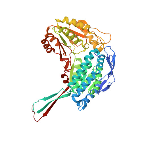
    Aldehyde dehydrogenase-2 (ALDH2) catalyzes the bioactivation of nitroglycerin (glyceryl trinitrate, GTN) in blood vessels, resulting in vasodilation by nitric oxide (NO) or a related species. Because the mechanism of this reaction is still unclear we determined the three-dimensional structures of wild-type (WT) ALDH2 and of a triple mutant of the protein that exhibits low denitration activity (E268Q/C301S/C303S) in complex with GTN. The structure of the triple mutant showed that GTN binds to the active site via polar contacts to the oxyanion hole and to residues 268 and 301 as well as by van der Waals interactions to hydrophobic residues of the catalytic pocket. The structure of the GTN-soaked wild-type protein revealed a thionitrate adduct to Cys-302 as the first reaction intermediate, which was also found by mass spectrometry (MS) experiments. In addition, the MS data identified sulfinic acid as the irreversibly inactivated enzyme species. Assuming that the structures of the triple mutant and wild-type ALDH2 reflect binding of GTN to the catalytic site and the first reaction step, respectively, superposition of the two structures indicates that denitration of GTN is initiated by nucleophilic attack of Cys-302 at one of the terminal nitrate groups, resulting in formation of the observed thionitrate intermediate and release of 1,2-glyceryl dinitrate. Our results shed light on the molecular mechanism of the GTN denitration reaction and provide useful information on the structural requirements for high affinity binding of organic nitrates to the catalytic site of ALDH2.


  • Organizational Affiliation
    • Department of Pharmacology and Toxicology, University of Graz, 8010 Graz, Austria.

Macromolecule Content 

  • Total Structure Weight: 441.27 kDa 
  • Atom Count: 33,739 
  • Modeled Residue Count: 3,948 
  • Deposited Residue Count: 4,000 
  • Unique protein chains: 1

Macromolecules

Find similar proteins by:|  3D Structure
Entity ID: 1
MoleculeChains  Sequence LengthOrganismDetailsImage
Aldehyde dehydrogenase, mitochondrial
A, B, C, D, E
A, B, C, D, E, F, G, H
500Homo sapiensMutation(s): 3 
Gene Names: ALDH2ALDM
EC: 1.2.1.3
UniProt & NIH Common Fund Data Resources
Find proteins for P05091 (Homo sapiens)
Explore P05091 
Go to UniProtKB:  P05091
PHAROS:  P05091
GTEx:  ENSG00000111275 
Entity Groups
Sequence Clusters30% Identity50% Identity70% Identity90% Identity95% Identity100% Identity
UniProt GroupP05091
Sequence Annotations
Expand
Reference Sequence

Small Molecules

Ligands 7 Unique
IDChains Name / Formula / InChI Key2D Diagram3D Interactions
NAD

Query on NAD


Download:Ideal Coordinates CCD File
EA [auth F],
J [auth A],
R [auth C],
W [auth D]
NICOTINAMIDE-ADENINE-DINUCLEOTIDE
C21 H27 N7 O14 P2
BAWFJGJZGIEFAR-NNYOXOHSSA-N
ADP

Query on ADP


Download:Ideal Coordinates CCD File
AA [auth E],
HA [auth G],
LA [auth H],
N [auth B]
ADENOSINE-5'-DIPHOSPHATE
C10 H15 N5 O10 P2
XTWYTFMLZFPYCI-KQYNXXCUSA-N
TNG

Query on TNG


Download:Ideal Coordinates CCD File
I [auth A]propane-1,2,3-triyl trinitrate
C3 H5 N3 O9
SNIOPGDIGTZGOP-UHFFFAOYSA-N
BTB

Query on BTB


Download:Ideal Coordinates CCD File
KA [auth G]2-[BIS-(2-HYDROXY-ETHYL)-AMINO]-2-HYDROXYMETHYL-PROPANE-1,3-DIOL
C8 H19 N O5
OWMVSZAMULFTJU-UHFFFAOYSA-N
URE

Query on URE


Download:Ideal Coordinates CCD File
DA [auth E]
L [auth A]
P [auth B]
T [auth C]
V [auth C]
DA [auth E],
L [auth A],
P [auth B],
T [auth C],
V [auth C],
Y [auth D]
UREA
C H4 N2 O
XSQUKJJJFZCRTK-UHFFFAOYSA-N
MG

Query on MG


Download:Ideal Coordinates CCD File
BA [auth E]
FA [auth F]
IA [auth G]
K [auth A]
MA [auth H]
BA [auth E],
FA [auth F],
IA [auth G],
K [auth A],
MA [auth H],
O [auth B],
S [auth C],
X [auth D]
MAGNESIUM ION
Mg
JLVVSXFLKOJNIY-UHFFFAOYSA-N
NA

Query on NA


Download:Ideal Coordinates CCD File
CA [auth E]
GA [auth F]
JA [auth G]
M [auth A]
NA [auth H]
CA [auth E],
GA [auth F],
JA [auth G],
M [auth A],
NA [auth H],
Q [auth B],
U [auth C],
Z [auth D]
SODIUM ION
Na
FKNQFGJONOIPTF-UHFFFAOYSA-N

Experimental Data & Validation

Experimental Data

  • Method: X-RAY DIFFRACTION
  • Resolution: 2.20 Å
  • R-Value Free:  0.167 (Depositor), 0.151 (DCC) 
  • R-Value Work:  0.132 (Depositor), 0.130 (DCC) 
  • R-Value Observed: 0.134 (Depositor) 
Space Group: P 1 21 1
Unit Cell:
Length ( Å )Angle ( ˚ )
a = 101.663α = 90
b = 175.916β = 95.01
c = 102.296γ = 90
Software Package:
Software NamePurpose
EDNAdata collection
PHASERphasing
PHENIXrefinement
XDSdata reduction
SCALAdata scaling

Structure Validation

View Full Validation Report


Ligand Structure Quality Assessment 


Entry History 

Deposition Data

Revision History  (Full details and data files)

  • Version 1.0: 2012-09-26
    Type: Initial release
  • Version 1.1: 2012-11-21
    Changes: Database references
  • Version 1.2: 2024-02-28
    Changes: Data collection, Database references, Derived calculations