4FLG | pdb_00004flg

HIV-1 protease mutant I47V complexed with reaction intermediate


Experimental Data Snapshot

  • Method: X-RAY DIFFRACTION
  • Resolution: 1.31 Å
  • R-Value Free: 
    0.180 (Depositor), 0.180 (DCC) 
  • R-Value Work: 
    0.142 (Depositor), 0.149 (DCC) 
  • R-Value Observed: 
    0.144 (Depositor) 

Starting Model: experimental
View more details

wwPDB Validation 3D Report Full Report

Validation slider image for 4FLG

This is version 1.3 of the entry. See complete history

Literature

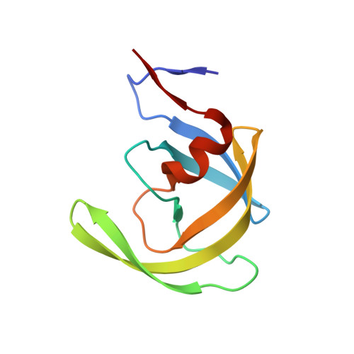
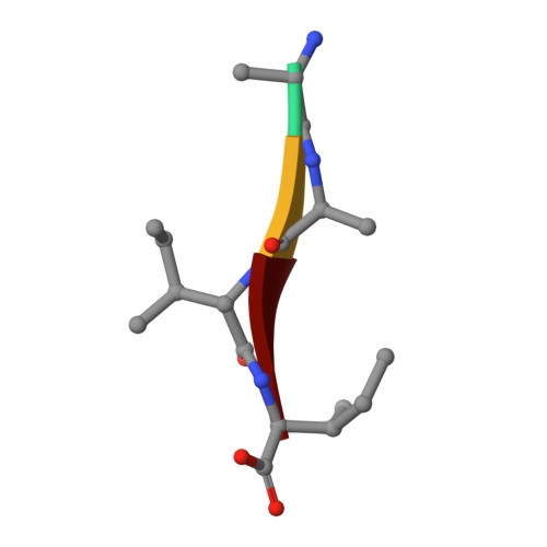
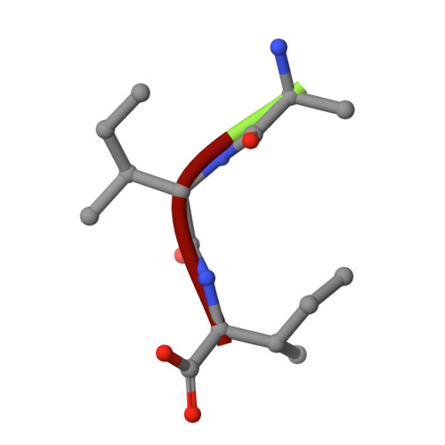
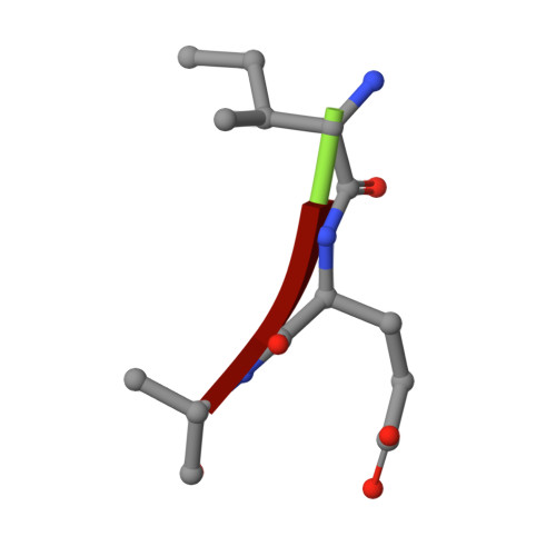
Capturing the Reaction Pathway in Near-Atomic-Resolution Crystal Structures of HIV-1 Protease.

Shen, C.H.Tie, Y.Yu, X.Wang, Y.F.Kovalevsky, A.Y.Harrison, R.W.Weber, I.T.

(2012) Biochemistry 51: 7726-7732

  • DOI: https://doi.org/10.1021/bi3008092
  • Primary Citation Related Structures: 
    4FL8, 4FLG, 4FM6

  • PubMed Abstract: 

    Snapshots of three consecutive steps in the proteolytic reaction of HIV-1 protease (PR) were obtained in crystal structures at resolutions of 1.2-1.4 Å. Structures of wild-type protease and two mutants (PR(V32I) and PR(I47V)) with V32I and I47V substitutions, which are common in drug resistance, reveal the gem-diol tetrahedral intermediate, the separating N- and C-terminal products, and the C-terminal product of an autoproteolytic peptide. These structures represent three stages in the reaction pathway and shed light on the reaction mechanism. The near-atomic-resolution geometric details include a short hydrogen bond between the intermediate and the outer carboxylate oxygen of one catalytic Asp25 that is conserved in all three structures. The two products in the complex with mutant PR(I47V) have a 2.2 Å separation of the amide and carboxyl carbon of the adjacent ends, suggesting partial cleavage prior to product release. The complex of mutant PR(V32I) with a single C-terminal product shows density for water molecules in the other half of the binding site, including a partial occupancy water molecule interacting with the product carboxylate end and the carbonyl oxygen of one conformation of Gly27, which suggests a potential role of Gly27 in recycling from the product complex to the ligand-free enzyme. These structural details at near-atomic resolution enhance our understanding of the reaction pathway and will assist in the design of mechanism-based inhibitors as antiviral agents.


  • Organizational Affiliation
    • Department of Biology, Molecular Basis of Disease Program, Georgia State University, Atlanta, Georgia 30303, United States.

Macromolecule Content 

  • Total Structure Weight: 23.31 kDa 
  • Atom Count: 1,868 
  • Modeled Residue Count: 208 
  • Deposited Residue Count: 208 
  • Unique protein chains: 4

Macromolecules

Find similar proteins by:|  3D Structure
Entity ID: 1
MoleculeChains  Sequence LengthOrganismDetailsImage
HIV-1 proteaseA [auth B],
B [auth A]
99Human immunodeficiency virus 1Mutation(s): 6 
EC: 3.4.23.16
UniProt
Find proteins for P03367 (Human immunodeficiency virus type 1 group M subtype B (isolate BRU/LAI))
Explore P03367 
Go to UniProtKB:  P03367
Entity Groups
Sequence Clusters30% Identity50% Identity70% Identity90% Identity95% Identity100% Identity
UniProt GroupP03367
Sequence Annotations
Expand
Reference Sequence
Find similar proteins by:  Sequence   |   3D Structure  
Entity ID: 2
MoleculeChains  Sequence LengthOrganismDetailsImage
HIV-1 protease, fragment4Human immunodeficiency virus 1Mutation(s): 0 
Gene Names: gag-pol
UniProt
Find proteins for P03367 (Human immunodeficiency virus type 1 group M subtype B (isolate BRU/LAI))
Explore P03367 
Go to UniProtKB:  P03367
Entity Groups
UniProt GroupP03367
Sequence Annotations
Expand
Reference Sequence
Find similar proteins by:  Sequence   |   3D Structure  
Entity ID: 3
MoleculeChains  Sequence LengthOrganismDetailsImage
HIV-1 protease, fragmentD [auth E]3Human immunodeficiency virus 1Mutation(s): 0 
UniProt
Find proteins for P03367 (Human immunodeficiency virus type 1 group M subtype B (isolate BRU/LAI))
Explore P03367 
Go to UniProtKB:  P03367
Entity Groups
UniProt GroupP03367
Sequence Annotations
Expand
Reference Sequence
Find similar proteins by:  Sequence   |   3D Structure  
Entity ID: 4
MoleculeChains  Sequence LengthOrganismDetailsImage
HIV-1 protease, fragmentE [auth F]3Human immunodeficiency virus 1Mutation(s): 0 
UniProt
Find proteins for P03367 (Human immunodeficiency virus type 1 group M subtype B (isolate BRU/LAI))
Explore P03367 
Go to UniProtKB:  P03367
Entity Groups
UniProt GroupP03367
Sequence Annotations
Expand
Reference Sequence

Small Molecules

Ligands 5 Unique
IDChains Name / Formula / InChI Key2D Diagram3D Interactions
GLU

Query on GLU


Download:Ideal Coordinates CCD File
F [auth B]GLUTAMIC ACID
C5 H9 N O4
WHUUTDBJXJRKMK-VKHMYHEASA-N
ILE

Query on ILE


Download:Ideal Coordinates CCD File
G [auth B]ISOLEUCINE
C6 H13 N O2
AGPKZVBTJJNPAG-WHFBIAKZSA-N
GOL

Query on GOL


Download:Ideal Coordinates CCD File
M [auth A],
N [auth A]
GLYCEROL
C3 H8 O3
PEDCQBHIVMGVHV-UHFFFAOYSA-N
CL

Query on CL


Download:Ideal Coordinates CCD File
I [auth B],
J [auth B],
K [auth B],
L [auth A]
CHLORIDE ION
Cl
VEXZGXHMUGYJMC-UHFFFAOYSA-M
NA

Query on NA


Download:Ideal Coordinates CCD File
H [auth B]SODIUM ION
Na
FKNQFGJONOIPTF-UHFFFAOYSA-N

Experimental Data & Validation

Experimental Data

  • Method: X-RAY DIFFRACTION
  • Resolution: 1.31 Å
  • R-Value Free:  0.180 (Depositor), 0.180 (DCC) 
  • R-Value Work:  0.142 (Depositor), 0.149 (DCC) 
  • R-Value Observed: 0.144 (Depositor) 
Space Group: P 21 21 2
Unit Cell:
Length ( Å )Angle ( ˚ )
a = 58.08α = 90
b = 86.3β = 90
c = 46.35γ = 90
Software Package:
Software NamePurpose
SERGUIdata collection
MOLREPphasing
SHELXL-97refinement
HKL-2000data reduction
HKL-2000data scaling

Structure Validation

View Full Validation Report


Entry History 

Deposition Data

Revision History  (Full details and data files)

  • Version 1.0: 2012-10-17
    Type: Initial release
  • Version 1.1: 2012-11-28
    Changes: Structure summary
  • Version 1.2: 2023-09-13
    Changes: Data collection, Database references, Derived calculations, Refinement description
  • Version 1.3: 2024-03-13
    Changes: Source and taxonomy