4FIO | pdb_00004fio

Crystal Structure of Methenyltetrahydromethanopterin Cyclohydrolase from Methanobrevibacter ruminantium


Experimental Data Snapshot

  • Method: X-RAY DIFFRACTION
  • Resolution: 1.37 Å
  • R-Value Free: 
    0.175 (Depositor), 0.163 (DCC) 
  • R-Value Work: 
    0.130 (Depositor), 0.124 (DCC) 
  • R-Value Observed: 
    0.132 (Depositor) 

Starting Model: experimental
View more details

wwPDB Validation 3D Report Full Report

Validation slider image for 4FIO

This is version 1.2 of the entry. See complete history

Literature

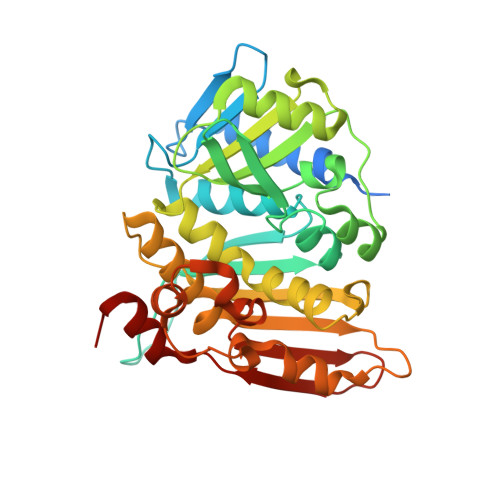
The crystal structure of methenyltetrahydromethanopterin cyclohydrolase from Methanobrevibacter ruminantium.

Carbone, V.Schofield, L.R.Beattie, A.K.Sutherland-Smith, A.J.Ronimus, R.S.

(2013) Proteins 81: 2064-2070

  • DOI: https://doi.org/10.1002/prot.24372
  • Primary Citation Related Structures: 
    4FIO

  • PubMed Abstract: 

    Methenyltetrahydromethanopterin cyclohydrolase (Mch) is involved in the methanogenesis pathway of archaea as a C1 unit carrier where N(5) -formyl-tetrahydromethanopterin is converted to methenyl-tetrahydromethanopterin. Mch from Methanobrevibacter ruminantium was cloned, purified, crystallized and its crystal structure solved at 1.37 Å resolution. A biologically active trimer, the enzyme is composed of two domains including an N-terminal domain of six α-helices encompassing a series of four β-sheets and a predominantly anti-parallel β-sheet at the C-terminus flanked on one side by α-helices. Sequence and structural alignments have helped identify residues involved in substrate binding and trimer formation.


  • Organizational Affiliation
    • AgResearch Limited, Grasslands Research Centre, Tennent Drive, Private Bag 11008, Palmerston North, 4442, New Zealand.

Macromolecule Content 

  • Total Structure Weight: 113.58 kDa 
  • Atom Count: 8,472 
  • Modeled Residue Count: 960 
  • Deposited Residue Count: 1,062 
  • Unique protein chains: 1

Macromolecules

Find similar proteins by:|  3D Structure
Entity ID: 1
MoleculeChains  Sequence LengthOrganismDetailsImage
Methenyltetrahydromethanopterin cyclohydrolase
A, B, C
354Methanobrevibacter ruminantium M1Mutation(s): 0 
Gene Names: mchmru_1619
EC: 3.5.4.27
UniProt
Find proteins for D3E4S5 (Methanobrevibacter ruminantium (strain ATCC 35063 / DSM 1093 / JCM 13430 / OCM 146 / M1))
Explore D3E4S5 
Go to UniProtKB:  D3E4S5
Entity Groups
Sequence Clusters30% Identity50% Identity70% Identity90% Identity95% Identity100% Identity
UniProt GroupD3E4S5
Sequence Annotations
Expand
Reference Sequence

Experimental Data & Validation

Experimental Data

  • Method: X-RAY DIFFRACTION
  • Resolution: 1.37 Å
  • R-Value Free:  0.175 (Depositor), 0.163 (DCC) 
  • R-Value Work:  0.130 (Depositor), 0.124 (DCC) 
  • R-Value Observed: 0.132 (Depositor) 
Space Group: P 1
Unit Cell:
Length ( Å )Angle ( ˚ )
a = 52.37α = 120.06
b = 74.855β = 100.01
c = 74.816γ = 98.28
Software Package:
Software NamePurpose
Blu-Icedata collection
MOLREPphasing
REFMACrefinement
XDSdata reduction
SCALAdata scaling

Structure Validation

View Full Validation Report


Entry History 

Deposition Data

Revision History  (Full details and data files)

  • Version 1.0: 2013-07-24
    Type: Initial release
  • Version 1.1: 2019-07-03
    Changes: Data collection, Database references
  • Version 1.2: 2023-09-13
    Changes: Data collection, Database references, Derived calculations, Refinement description