4FGO

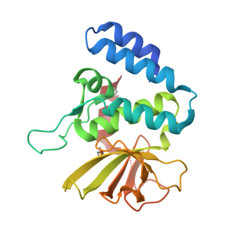
Legionella pneumophila LapG (calcium-bound)


Experimental Data Snapshot

  • Method: X-RAY DIFFRACTION
  • Resolution: 1.90 Å
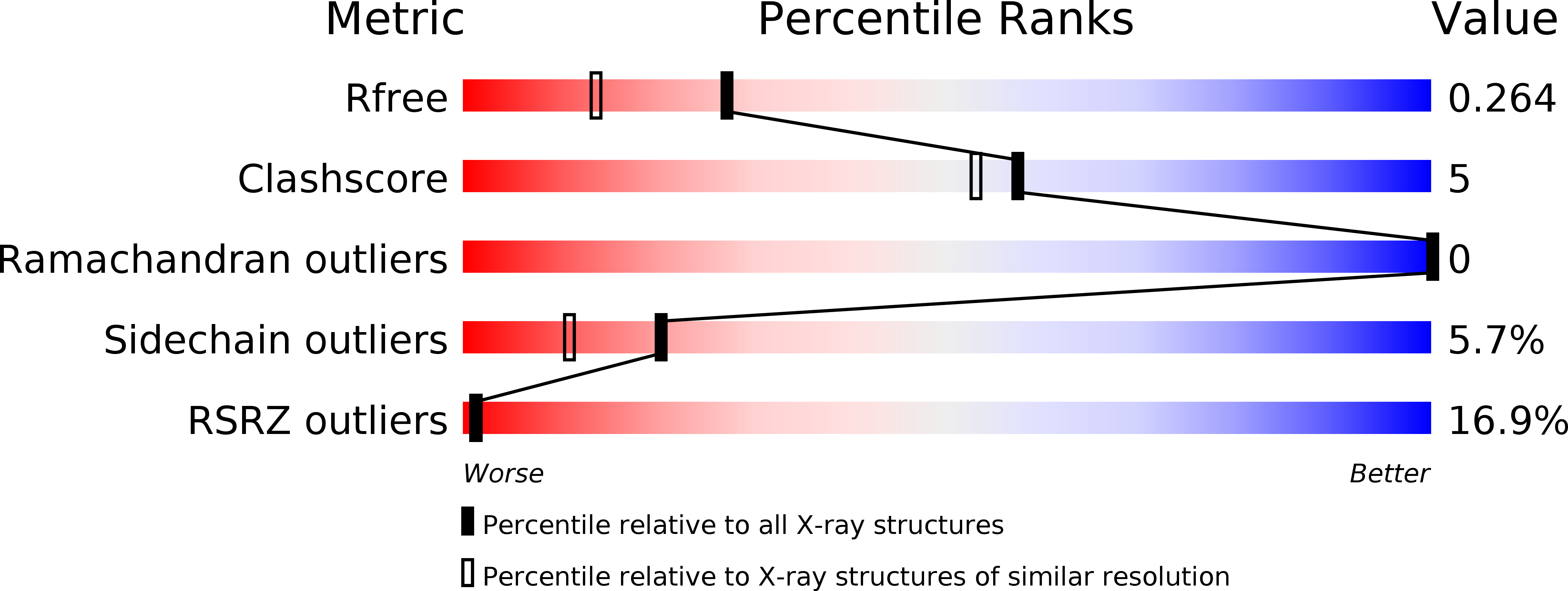
  • R-Value Free: 0.267 
  • R-Value Work: 0.259 
  • R-Value Observed: 0.260 

wwPDB Validation   3D Report Full Report


This is version 1.2 of the entry. See complete history


Literature

Structural characterization of a conserved, calcium-dependent periplasmic protease from Legionella pneumophila.

Chatterjee, D.Boyd, C.D.O'Toole, G.A.Sondermann, H.

(2012) J Bacteriol 194: 4415-4425

  • DOI: https://doi.org/10.1128/JB.00640-12
  • Primary Citation of Related Structures:  
    4FGO, 4FGP, 4FGQ

  • PubMed Abstract: 

    The bacterial dinucleotide second messenger c-di-GMP has emerged as a central molecule in regulating bacterial behavior, including motility and biofilm formation. Proteins for the synthesis and degradation of c-di-GMP and effectors for its signal transmission are widely used in the bacterial domain. Previous work established the GGDEF-EAL domain-containing receptor LapD as a central switch in Pseudomonas fluorescens cell adhesion. LapD senses c-di-GMP inside the cytosol and relays this signal to the outside by the differential recruitment of the periplasmic protease LapG. Here we identify the core components of an orthologous system in Legionella pneumophila. Despite only moderate sequence conservation at the protein level, key features concerning the regulation of LapG are retained. The output domain of the LapD-like receptor from L. pneumophila, CdgS9, binds the LapG ortholog involving a strictly conserved surface tryptophan residue. While the endogenous substrate for L. pneumophila LapG is unknown, the enzyme processed the corresponding P. fluorescens substrate, indicating a common catalytic mechanism and substrate recognition. Crystal structures of L. pneumophila LapG provide the first atomic models of bacterial proteases of the DUF920 family and reveal a conserved calcium-binding site important for LapG function.


  • Organizational Affiliation

    Department of Molecular Medicine, College of Veterinary Medicine, Cornell University, Ithaca, New York, USA.


Macromolecules
Find similar proteins by:  (by identity cutoff)  |  3D Structure
Entity ID: 1
MoleculeChains Sequence LengthOrganismDetailsImage
Periplasmic protein193Legionella pneumophila subsp. pneumophila str. Philadelphia 1Mutation(s): 0 
Gene Names: lpg0828
UniProt
Find proteins for Q5ZXA4 (Legionella pneumophila subsp. pneumophila (strain Philadelphia 1 / ATCC 33152 / DSM 7513))
Explore Q5ZXA4 
Go to UniProtKB:  Q5ZXA4
Entity Groups  
Sequence Clusters30% Identity50% Identity70% Identity90% Identity95% Identity100% Identity
UniProt GroupQ5ZXA4
Sequence Annotations
Expand
  • Reference Sequence
Small Molecules
Ligands 1 Unique
IDChains Name / Formula / InChI Key2D Diagram3D Interactions
CA
Query on CA

Download Ideal Coordinates CCD File 
B [auth A]CALCIUM ION
Ca
BHPQYMZQTOCNFJ-UHFFFAOYSA-N
Modified Residues  1 Unique
IDChains TypeFormula2D DiagramParent
MSE
Query on MSE
A
L-PEPTIDE LINKINGC5 H11 N O2 SeMET
Experimental Data & Validation

Experimental Data

  • Method: X-RAY DIFFRACTION
  • Resolution: 1.90 Å
  • R-Value Free: 0.267 
  • R-Value Work: 0.259 
  • R-Value Observed: 0.260 
  • Space Group: P 43 21 2
Unit Cell:
Length ( Å )Angle ( ˚ )
a = 59.951α = 90
b = 59.951β = 90
c = 110.379γ = 90
Software Package:
Software NamePurpose
ADSCdata collection
PHENIXmodel building
PHENIXrefinement
HKL-2000data reduction
HKL-2000data scaling
PHENIXphasing

Structure Validation

View Full Validation Report


Entry History 

Deposition Data

Revision History  (Full details and data files)

  • Version 1.0: 2012-06-20
    Type: Initial release
  • Version 1.1: 2013-01-09
    Changes: Database references
  • Version 1.2: 2024-10-16
    Changes: Data collection, Database references, Derived calculations, Structure summary