4FCT | pdb_00004fct

Crystal structure of the C-terminal domain of ClpB


Experimental Data Snapshot

  • Method: X-RAY DIFFRACTION
  • Resolution: 4.00 Å
  • R-Value Free: 
    0.297 (Depositor), 0.316 (DCC) 
  • R-Value Work: 
    0.285 (Depositor), 0.284 (DCC) 
  • R-Value Observed: 
    0.286 (Depositor) 

wwPDB Validation 3D Report Full Report

Validation slider image for 4FCT

This is version 1.2 of the entry. See complete history

Literature

Structural basis for intersubunit signaling in a protein disaggregating machine.

Biter, A.B.Lee, S.Sung, N.Tsai, F.T.

(2012) Proc Natl Acad Sci U S A 109: 12515-12520

  • DOI: https://doi.org/10.1073/pnas.1207040109
  • Primary Citation Related Structures: 
    4FCT, 4FCV, 4FCW, 4FD2

  • PubMed Abstract: 

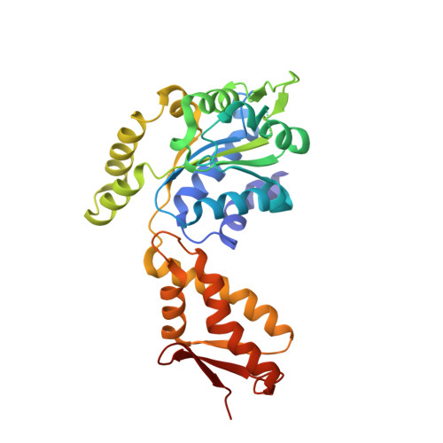
    ClpB is a ring-forming, ATP-dependent protein disaggregase that cooperates with the cognate Hsp70 system to recover functional protein from aggregates. How ClpB harnesses the energy of ATP binding and hydrolysis to facilitate the mechanical unfolding of previously aggregated, stress-damaged proteins remains unclear. Here, we present crystal structures of the ClpB D2 domain in the nucleotide-bound and -free states, and the fitted cryoEM structure of the D2 hexamer ring, which provide a structural understanding of the ATP power stroke that drives protein translocation through the ClpB hexamer. We demonstrate that the conformation of the substrate-translocating pore loop is coupled to the nucleotide state of the cis subunit, which is transmitted to the neighboring subunit via a conserved but structurally distinct intersubunit-signaling pathway common to diverse AAA+ machines. Furthermore, we found that an engineered, disulfide cross-linked ClpB hexamer is fully functional biochemically, suggesting that ClpB deoligomerization is not required for protein disaggregation.


  • Organizational Affiliation
    • Program in Structural and Computational Biology and Molecular Biophysics, Baylor College of Medicine, Houston, TX 77030, USA.

Macromolecule Content 

  • Total Structure Weight: 34.93 kDa 
  • Atom Count: 2,348 
  • Modeled Residue Count: 292 
  • Deposited Residue Count: 308 
  • Unique protein chains: 1

Macromolecules

Find similar proteins by:|  3D Structure
Entity ID: 1
MoleculeChains  Sequence LengthOrganismDetailsImage
Chaperone protein ClpB308Thermus thermophilus HB8Mutation(s): 0 
Gene Names: clpBTTHA1487
UniProt
Find proteins for Q9RA63 (Thermus thermophilus (strain ATCC 27634 / DSM 579 / HB8))
Explore Q9RA63 
Go to UniProtKB:  Q9RA63
Entity Groups
Sequence Clusters30% Identity50% Identity70% Identity90% Identity95% Identity100% Identity
UniProt GroupQ9RA63
Sequence Annotations
Expand
Reference Sequence

Experimental Data & Validation

Experimental Data

  • Method: X-RAY DIFFRACTION
  • Resolution: 4.00 Å
  • R-Value Free:  0.297 (Depositor), 0.316 (DCC) 
  • R-Value Work:  0.285 (Depositor), 0.284 (DCC) 
  • R-Value Observed: 0.286 (Depositor) 
Space Group: P 65
Unit Cell:
Length ( Å )Angle ( ˚ )
a = 56.22α = 90
b = 56.22β = 90
c = 198.71γ = 120
Software Package:
Software NamePurpose
REFMACrefinement
PDB_EXTRACTdata extraction

Structure Validation

View Full Validation Report


Entry History 

Deposition Data

Revision History  (Full details and data files)

  • Version 1.0: 2012-07-18
    Type: Initial release
  • Version 1.1: 2012-08-22
    Changes: Database references
  • Version 1.2: 2024-02-28
    Changes: Data collection, Database references