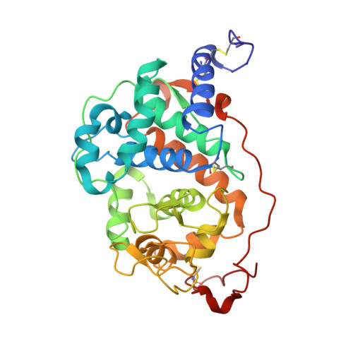
4FCS

The crystal structures of several mutants of pleurotus eryngii versatile peroxidase


Experimental Data Snapshot

  • Method: X-RAY DIFFRACTION
  • Resolution: 1.50 Å
  • R-Value Free: 0.176 
  • R-Value Work: 0.150 
  • R-Value Observed: 0.151 

Starting Model: experimental
View more details

wwPDB Validation   3D Report Full Report


Ligand Structure Quality Assessment 


This is version 1.3 of the entry. See complete history


Literature

Two Oxidation Sites for Low Redox Potential Substrates: A DIRECTED MUTAGENESIS, KINETIC, AND CRYSTALLOGRAPHIC STUDY ON PLEUROTUS ERYNGII VERSATILE PEROXIDASE.

Morales, M.Mate, M.J.Romero, A.Martinez, M.J.Martinez, A.T.Ruiz-Duenas, F.J.

(2012) J Biol Chem 287: 41053-41067

  • DOI: https://doi.org/10.1074/jbc.M112.405548
  • Primary Citation of Related Structures:  
    4FCN, 4FCS, 4FDQ, 4FEF, 4G05

  • PubMed Abstract: 

    Versatile peroxidase shares with manganese peroxidase and lignin peroxidase the ability to oxidize Mn(2+) and high redox potential aromatic compounds, respectively. Moreover, it is also able to oxidize phenols (and low redox potential dyes) at two catalytic sites, as shown by biphasic kinetics. A high efficiency site (with 2,6-dimethoxyphenol and p-hydroquinone catalytic efficiencies of ∼70 and ∼700 s(-1) mM(-1), respectively) was localized at the same exposed Trp-164 responsible for high redox potential substrate oxidation (as shown by activity loss in the W164S variant). The second site, characterized by low catalytic efficiency (∼3 and ∼50 s(-1) mM(-1) for 2,6-dimethoxyphenol and p-hydroquinone, respectively) was localized at the main heme access channel. Steady-state and transient-state kinetics for oxidation of phenols and dyes at the latter site were improved when side chains of residues forming the heme channel edge were removed in single and multiple variants. Among them, the E140G/K176G, E140G/P141G/K176G, and E140G/W164S/K176G variants attained catalytic efficiencies for oxidation of 2,2'-azino-bis(3-ethylbenzothiazoline-6-sulfonate) at the heme channel similar to those of the exposed tryptophan site. The heme channel enlargement shown by x-ray diffraction of the E140G, P141G, K176G, and E140G/K176G variants would allow a better substrate accommodation near the heme, as revealed by the up to 26-fold lower K(m) values (compared with native VP). The resulting interactions were shown by the x-ray structure of the E140G-guaiacol complex, which includes two H-bonds of the substrate with Arg-43 and Pro-139 in the distal heme pocket (at the end of the heme channel) and several hydrophobic interactions with other residues and the heme cofactor.


  • Organizational Affiliation

    Centro de Investigaciones Biológicas (CIB), CSIC, Ramiro de Maeztu 9, E-28040 Madrid, Spain.


Macromolecules
Find similar proteins by:  (by identity cutoff)  |  3D Structure
Entity ID: 1
MoleculeChains Sequence LengthOrganismDetailsImage
Versatile peroxidase VPL2315Pleurotus eryngiiMutation(s): 2 
Gene Names: vpl2
EC: 1.11.1.16
UniProt
Find proteins for O94753 (Pleurotus eryngii)
Explore O94753 
Go to UniProtKB:  O94753
Entity Groups  
Sequence Clusters30% Identity50% Identity70% Identity90% Identity95% Identity100% Identity
UniProt GroupO94753
Sequence Annotations
Expand
  • Reference Sequence
Small Molecules
Ligands 4 Unique
IDChains Name / Formula / InChI Key2D Diagram3D Interactions
HEM
Query on HEM

Download Ideal Coordinates CCD File 
B [auth A]PROTOPORPHYRIN IX CONTAINING FE
C34 H32 Fe N4 O4
KABFMIBPWCXCRK-RGGAHWMASA-L
SO4
Query on SO4

Download Ideal Coordinates CCD File 
E [auth A]SULFATE ION
O4 S
QAOWNCQODCNURD-UHFFFAOYSA-L
GOL
Query on GOL

Download Ideal Coordinates CCD File 
F [auth A],
G [auth A]
GLYCEROL
C3 H8 O3
PEDCQBHIVMGVHV-UHFFFAOYSA-N
CA
Query on CA

Download Ideal Coordinates CCD File 
C [auth A],
D [auth A]
CALCIUM ION
Ca
BHPQYMZQTOCNFJ-UHFFFAOYSA-N
Experimental Data & Validation

Experimental Data

  • Method: X-RAY DIFFRACTION
  • Resolution: 1.50 Å
  • R-Value Free: 0.176 
  • R-Value Work: 0.150 
  • R-Value Observed: 0.151 
  • Space Group: I 41
Unit Cell:
Length ( Å )Angle ( ˚ )
a = 96.271α = 90
b = 96.271β = 90
c = 98.907γ = 90
Software Package:
Software NamePurpose
DNAdata collection
MOLREPphasing
REFMACrefinement
XDSdata reduction
SCALAdata scaling

Structure Validation

View Full Validation Report


Ligand Structure Quality Assessment 


Entry History 

Deposition Data

Revision History  (Full details and data files)

  • Version 1.0: 2012-10-24
    Type: Initial release
  • Version 1.1: 2012-12-19
    Changes: Database references
  • Version 1.2: 2023-09-13
    Changes: Data collection, Database references, Derived calculations, Refinement description
  • Version 1.3: 2024-11-06
    Changes: Structure summary