4FCH | pdb_00004fch

Crystal Structure SusE from Bacteroides thetaiotaomicron with maltoheptaose


Experimental Data Snapshot

  • Method: X-RAY DIFFRACTION
  • Resolution: 1.30 Å
  • R-Value Free: 
    0.177 (Depositor), 0.186 (DCC) 
  • R-Value Work: 
    0.164 (Depositor), 0.177 (DCC) 
  • R-Value Observed: 
    0.165 (Depositor) 

Starting Model: experimental
View more details

wwPDB Validation 3D Report Full Report

Validation slider image for 4FCH

This is version 2.2 of the entry. See complete history

Literature

Multidomain Carbohydrate-binding Proteins Involved in Bacteroides thetaiotaomicron Starch Metabolism.

Cameron, E.A.Maynard, M.A.Smith, C.J.Smith, T.J.Koropatkin, N.M.Martens, E.C.

(2012) J Biological Chem 287: 34614-34625

  • DOI: https://doi.org/10.1074/jbc.M112.397380
  • Primary Citation Related Structures: 
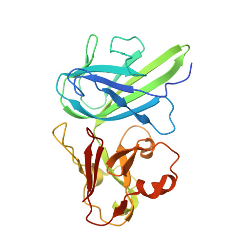
    4FCH, 4FE9, 4FEM

  • PubMed Abstract: 

    Human colonic bacteria are necessary for the digestion of many dietary polysaccharides. The intestinal symbiont Bacteroides thetaiotaomicron uses five outer membrane proteins to bind and degrade starch. Here, we report the x-ray crystallographic structures of SusE and SusF, two outer membrane proteins composed of tandem starch specific carbohydrate-binding modules (CBMs) with no enzymatic activity. Examination of the two CBMs in SusE and three CBMs in SusF reveals subtle differences in the way each binds starch and is reflected in their K(d) values for both high molecular weight starch and small maltooligosaccharides. Thus, each site seems to have a unique starch preference that may enable these proteins to interact with different regions of starch or its breakdown products. Proteins similar to SusE and SusF are encoded in many other polysaccharide utilization loci that are possessed by human gut bacteria in the phylum Bacteroidetes. Thus, these proteins are likely to play an important role in carbohydrate metabolism in these abundant symbiotic species. Understanding structural changes that diversify and adapt related proteins in the human gut microbial community will be critical to understanding the detailed mechanistic roles that they perform in the complex digestive ecosystem.


  • Organizational Affiliation
    • Department of Microbiology and Immunology, University of Michigan Medical School, Ann Arbor, Michigan 48109, USA.

Macromolecule Content 

  • Total Structure Weight: 51.67 kDa 
  • Atom Count: 4,142 
  • Modeled Residue Count: 426 
  • Deposited Residue Count: 442 
  • Unique protein chains: 1

Macromolecules

Find similar proteins by:|  3D Structure
Entity ID: 1
MoleculeChains  Sequence LengthOrganismDetailsImage
Outer membrane protein SusE
A, B
221Bacteroides thetaiotaomicron VPI-5482Mutation(s): 0 
Gene Names: BT_3700susE
UniProt
Find proteins for G8JZT0 (Bacteroides thetaiotaomicron (strain ATCC 29148 / DSM 2079 / JCM 5827 / CCUG 10774 / NCTC 10582 / VPI-5482 / E50))
Explore G8JZT0 
Go to UniProtKB:  G8JZT0
Entity Groups
Sequence Clusters30% Identity50% Identity70% Identity90% Identity95% Identity100% Identity
UniProt GroupG8JZT0
Sequence Annotations
Expand
Reference Sequence

Oligosaccharides

Help  
Entity ID: 2
MoleculeChains Length2D Diagram GlycosylationD Interactions
alpha-D-glucopyranose-(1-4)-alpha-D-glucopyranose-(1-4)-alpha-D-glucopyranose-(1-4)-alpha-D-glucopyranose-(1-4)-alpha-D-glucopyranose-(1-4)-alpha-D-glucopyranose-(1-4)-alpha-D-glucopyranose
C
7N/A
Glycosylation Resources
GlyTouCan: G41288IQ
GlyCosmos: G41288IQ
GlyGen: G41288IQ

Small Molecules

Ligands 2 Unique
IDChains Name / Formula / InChI Key2D Diagram3D Interactions
SO4

Query on SO4


Download:Ideal Coordinates CCD File
D [auth A],
E [auth A],
K [auth B],
L [auth B]
SULFATE ION
O4 S
QAOWNCQODCNURD-UHFFFAOYSA-L
EDO

Query on EDO


Download:Ideal Coordinates CCD File
F [auth A]
G [auth A]
H [auth A]
I [auth A]
J [auth A]
F [auth A],
G [auth A],
H [auth A],
I [auth A],
J [auth A],
M [auth B],
N [auth B],
O [auth B],
P [auth B],
Q [auth B]
1,2-ETHANEDIOL
C2 H6 O2
LYCAIKOWRPUZTN-UHFFFAOYSA-N

Experimental Data & Validation

Experimental Data

  • Method: X-RAY DIFFRACTION
  • Resolution: 1.30 Å
  • R-Value Free:  0.177 (Depositor), 0.186 (DCC) 
  • R-Value Work:  0.164 (Depositor), 0.177 (DCC) 
  • R-Value Observed: 0.165 (Depositor) 
Space Group: P 21 21 21
Unit Cell:
Length ( Å )Angle ( ˚ )
a = 58.735α = 90
b = 63.914β = 90
c = 156.828γ = 90
Software Package:
Software NamePurpose
MD2data collection
SOLVEphasing
REFMACrefinement
HKL-2000data reduction
HKL-2000data scaling

Structure Validation

View Full Validation Report


Entry History 

Deposition Data

Revision History  (Full details and data files)

  • Version 1.0: 2012-08-29
    Type: Initial release
  • Version 1.1: 2012-09-05
    Changes: Database references
  • Version 1.2: 2012-10-24
    Changes: Database references
  • Version 2.0: 2020-07-29
    Type: Remediation
    Reason: Carbohydrate remediation
    Changes: Atomic model, Data collection, Database references, Derived calculations, Structure summary
  • Version 2.1: 2024-02-28
    Changes: Data collection, Database references, Structure summary
  • Version 2.2: 2024-04-03
    Changes: Refinement description