4FA8 | pdb_00004fa8

Multi-pronged modulation of cytokine signaling


Experimental Data Snapshot

  • Method: X-RAY DIFFRACTION
  • Resolution: 2.20 Å
  • R-Value Free: 
    0.260 (Depositor), 0.262 (DCC) 
  • R-Value Work: 
    0.222 (Depositor), 0.222 (DCC) 
  • R-Value Observed: 
    0.224 (Depositor) 

wwPDB Validation 3D Report Full Report

Validation slider image for 4FA8

Ligand Structure Quality Assessment 


This is version 2.1 of the entry. See complete history

Literature

Multipronged attenuation of macrophage-colony stimulating factor signaling by Epstein-Barr virus BARF1.

Shim, A.H.Chang, R.A.Chen, X.Longnecker, R.He, X.

(2012) Proc Natl Acad Sci U S A 109: 12962-12967

  • DOI: https://doi.org/10.1073/pnas.1205309109
  • Primary Citation Related Structures: 
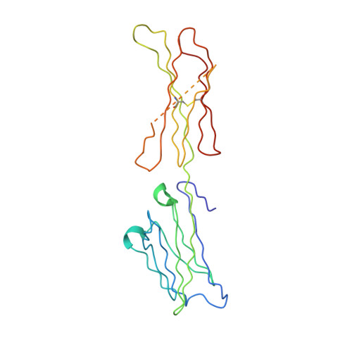
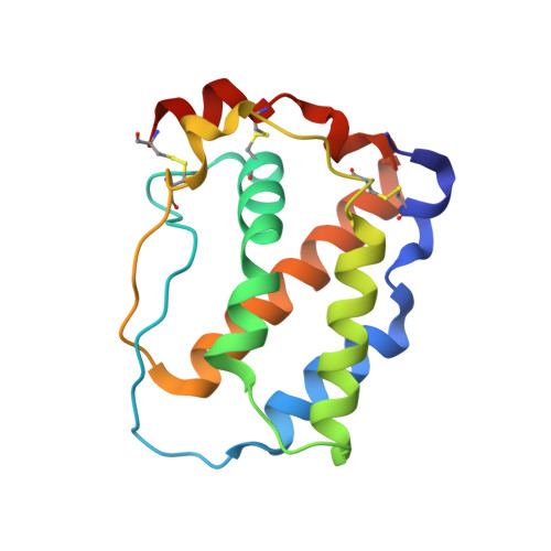
    4FA8

  • PubMed Abstract: 

    The ubiquitous EBV causes infectious mononucleosis and is associated with several types of cancers. The EBV genome encodes an early gene product, BARF1, which contributes to pathogenesis, potentially through growth-altering and immune-modulating activities, but the mechanisms for such activities are poorly understood. We have determined the crystal structure of BARF1 in complex with human macrophage-colony stimulating factor (M-CSF), a hematopoietic cytokine with pleiotropic functions in development and immune response. BARF1 and M-CSF form a high-affinity, stable, ring-like complex in both solution and the crystal, with a BARF1 hexameric ring surrounded by three M-CSF dimers in triangular array. The binding of BARF1 to M-CSF dramatically reduces but does not completely abolish M-CSF binding and signaling through its cognate receptor FMS. A three-pronged down-regulation mechanism is proposed to explain the biological effect of BARF1 on M-CSF:FMS signaling. These prongs entail control of the circulating and effective local M-CSF concentration, perturbation of the receptor-binding surface of M-CSF, and imposition of an unfavorable global orientation of the M-CSF dimer. Each prong may reduce M-CSF:FMS signaling to a limited extent but in combination may alter M-CSF:FMS signaling dramatically. The downregulating mechanism of BARF1 underlines a viral modulation strategy, and provides a basis for understanding EBV pathogenesis.


  • Organizational Affiliation
    • Department of Molecular Pharmacology and Biological Chemistry, Feinberg School of Medicine, Northwestern University, Chicago, IL 60611, USA.

Macromolecule Content 

  • Total Structure Weight: 121.97 kDa 
  • Atom Count: 8,676 
  • Modeled Residue Count: 1,003 
  • Deposited Residue Count: 1,050 
  • Unique protein chains: 2

Macromolecules

Find similar proteins by:|  3D Structure
Entity ID: 1
MoleculeChains  Sequence LengthOrganismDetailsImage
Secreted protein BARF1A,
B,
C [auth D]
203human gammaherpesvirus 4Mutation(s): 0 
Gene Names: BARF1
UniProt
Find proteins for P0CW72 (Epstein-Barr virus (strain GD1))
Explore P0CW72 
Go to UniProtKB:  P0CW72
Entity Groups
Sequence Clusters30% Identity50% Identity70% Identity90% Identity95% Identity100% Identity
UniProt GroupP0CW72
Glycosylation
Glycosylation Sites: 1
Sequence Annotations
Expand
Reference Sequence
Find similar proteins by:|  3D Structure
Entity ID: 2
MoleculeChains  Sequence LengthOrganismDetailsImage
Macrophage colony-stimulating factor 1D [auth E],
E [auth F],
F [auth G]
147Homo sapiensMutation(s): 0 
Gene Names: CSF1
UniProt & NIH Common Fund Data Resources
Find proteins for P09603 (Homo sapiens)
Explore P09603 
Go to UniProtKB:  P09603
PHAROS:  P09603
GTEx:  ENSG00000184371 
Entity Groups
Sequence Clusters30% Identity50% Identity70% Identity90% Identity95% Identity100% Identity
UniProt GroupP09603
Glycosylation
Glycosylation Sites: 1Go to GlyGen: P09603-1
Sequence Annotations
Expand
Reference Sequence

Oligosaccharides

Help  
Entity ID: 3
MoleculeChains Length2D Diagram GlycosylationD Interactions
beta-D-mannopyranose-(1-4)-2-acetamido-2-deoxy-beta-D-glucopyranose-(1-4)-2-acetamido-2-deoxy-beta-D-glucopyranoseG [auth C],
H,
I
3N-Glycosylation
Glycosylation Resources
GlyTouCan: G15407YE
GlyCosmos: G15407YE
GlyGen: G15407YE

Experimental Data & Validation

Experimental Data

  • Method: X-RAY DIFFRACTION
  • Resolution: 2.20 Å
  • R-Value Free:  0.260 (Depositor), 0.262 (DCC) 
  • R-Value Work:  0.222 (Depositor), 0.222 (DCC) 
  • R-Value Observed: 0.224 (Depositor) 
Space Group: C 1 2 1
Unit Cell:
Length ( Å )Angle ( ˚ )
a = 199.554α = 90
b = 162.701β = 95.87
c = 57.337γ = 90
Software Package:
Software NamePurpose
HKL-2000data collection
PHASERphasing
REFMACrefinement
HKL-2000data reduction
HKL-2000data scaling

Structure Validation

View Full Validation Report


Ligand Structure Quality Assessment 


Entry History 

Deposition Data

Revision History  (Full details and data files)

  • Version 1.0: 2012-08-29
    Type: Initial release
  • Version 2.0: 2020-07-29
    Type: Remediation
    Reason: Carbohydrate remediation
    Changes: Advisory, Atomic model, Data collection, Database references, Derived calculations, Structure summary
  • Version 2.1: 2024-11-20
    Changes: Data collection, Database references, Refinement description, Structure summary